Saltar al contenido
Merck
Todas las fotos(4)

Key Documents

HPA020433

Sigma-Aldrich

Anti-PLS3 antibody produced in rabbit

enhanced validation

Prestige Antibodies® Powered by Atlas Antibodies, affinity isolated antibody, buffered aqueous glycerol solution

Sinónimos:

Anti-T-plastin, Anti-plastin 3

Iniciar sesiónpara Ver la Fijación de precios por contrato y de la organización


About This Item

UNSPSC Code:
12352203
Human Protein Atlas Number:
NACRES:
NA.41

biological source

rabbit

conjugate

unconjugated

antibody form

affinity isolated antibody

antibody product type

primary antibodies

clone

polyclonal

product line

Prestige Antibodies® Powered by Atlas Antibodies

form

buffered aqueous glycerol solution

species reactivity

human

enhanced validation

orthogonal RNAseq
Learn more about Antibody Enhanced Validation

technique(s)

immunoblotting: 0.04-0.4 μg/mL
immunohistochemistry: 1:50-1:200

immunogen sequence

PAKFSLVGIGGQDLNDGNQTLTLALVWQLMRRYTLNVLEDLGDGQKANDDIIVNWVNRTLSEAGKSTSIQSFKDKTISSSLAVVDLIDAIQPGCINYDLVKSGNLTEDDKHNNAKYAVSMARRIGARVYALPEDLVEVKPGAPHR

UniProt accession no.

shipped in

wet ice

storage temp.

−20°C

target post-translational modification

unmodified

Gene Information

human ... PLS3(5358)

General description

Plastin 3 (PLS3) is expressed in epithelial cells. The gene encoding it is localized on human chromosome Xq23.

Immunogen

plastin 3 recombinant protein epitope signature tag (PrEST)

Application

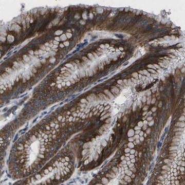
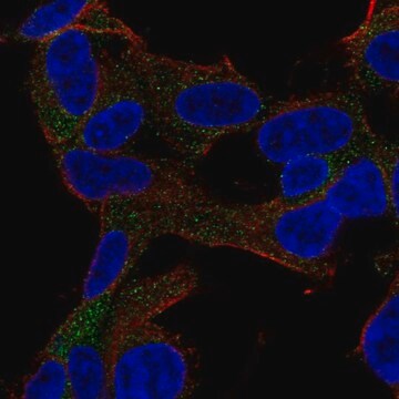
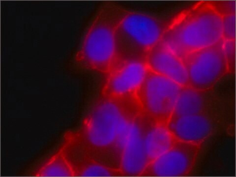
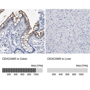
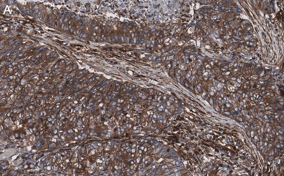
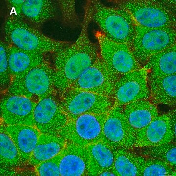
All Prestige Antibodies Powered by Atlas Antibodies are developed and validated by the Human Protein Atlas (HPA) project and as a result, are supported by the most extensive characterization in the industry.

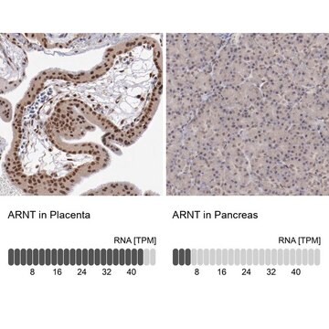
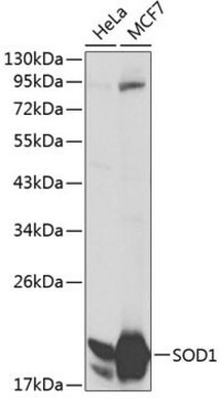
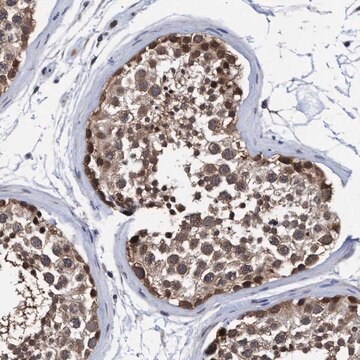
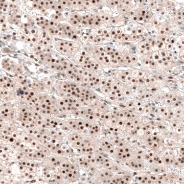
The Human Protein Atlas project can be subdivided into three efforts: Human Tissue Atlas, Cancer Atlas, and Human Cell Atlas. The antibodies that have been generated in support of the Tissue and Cancer Atlas projects have been tested by immunohistochemistry against hundreds of normal and disease tissues and through the recent efforts of the Human Cell Atlas project, many have been characterized by immunofluorescence to map the human proteome not only at the tissue level but now at the subcellular level. These images and the collection of this vast data set can be viewed on the Human Protein Atlas (HPA) site by clicking on the Image Gallery link. We also provide Prestige Antibodies® protocols and other useful information.

Biochem/physiol Actions

Plastin 3 (PLS3) imparts the bundled nature to actin filaments and cross-links to them, thereby helping in cell motility. In circulating tumor cells, it stimulates epithelial-mesenchymal transition (EMT) through the transforming growth factor-β (TGF-β) pathway. Mutation in the PLS3 gene leads to X-linked low-turnover osteoporosis and the protein has been found to be expressed in breast cancer.

Features and Benefits

Prestige Antibodies® are highly characterized and extensively validated antibodies with the added benefit of all available characterization data for each target being accessible via the Human Protein Atlas portal linked just below the product name at the top of this page. The uniqueness and low cross-reactivity of the Prestige Antibodies® to other proteins are due to a thorough selection of antigen regions, affinity purification, and stringent selection. Prestige antigen controls are available for every corresponding Prestige Antibody and can be found in the linkage section.

Every Prestige Antibody is tested in the following ways:
  • IHC tissue array of 44 normal human tissues and 20 of the most common cancer type tissues.
  • Protein array of 364 human recombinant protein fragments.

Linkage

Corresponding Antigen APREST82421

Physical form

Solution in phosphate buffered saline, pH 7.2, containing 40% glycerol and 0.02% sodium azide.

Legal Information

Prestige Antibodies is a registered trademark of Merck KGaA, Darmstadt, Germany

Disclaimer

Unless otherwise stated in our catalog or other company documentation accompanying the product(s), our products are intended for research use only and are not to be used for any other purpose, which includes but is not limited to, unauthorized commercial uses, in vitro diagnostic uses, ex vivo or in vivo therapeutic uses or any type of consumption or application to humans or animals.

¿No encuentra el producto adecuado?  

Pruebe nuestro Herramienta de selección de productos.

Storage Class

10 - Combustible liquids

wgk_germany

WGK 1

flash_point_f

Not applicable

flash_point_c

Not applicable


Certificados de análisis (COA)

Busque Certificados de análisis (COA) introduciendo el número de lote del producto. Los números de lote se encuentran en la etiqueta del producto después de las palabras «Lot» o «Batch»

¿Ya tiene este producto?

Encuentre la documentación para los productos que ha comprado recientemente en la Biblioteca de documentos.

Visite la Librería de documentos

Christine M Laine et al.
Journal of bone and mineral research : the official journal of the American Society for Bone and Mineral Research, 30(3), 510-518 (2014-09-12)
Genetic factors play an important role in the development of osteoporosis. Several monogenic forms of osteoporosis have been recognized, most recently an X-chromosomal form resulting from mutations in the gene encoding plastin 3 (PLS3). PLS3 is a protein involved in
Christine L Jones et al.
The Journal of investigative dermatology, 132(8), 2042-2049 (2012-04-13)
T-plastin (PLS3) is an actin-bundling protein normally expressed in epithelial cells but absent in cells of hematopoietic origin. Aberrant PLS3 expression has been demonstrated in lymphocytes from Sézary syndrome (SS) patients and has been proposed as a biomarker for SS;
Cécilia Brun et al.
PloS one, 9(9), e104700-e104700 (2014-09-17)
Cutaneous wound healing requires keratinocyte proliferation, migration and differentiation to restore the barrier function of the skin. The calcineurin/nuclear factor of activated-T-cell (NFAT) signaling pathway has been recently shown to be involved in keratinocyte growth, differentiation and migration. It is
Nan Li et al.
FASEB journal : official publication of the Federation of American Societies for Experimental Biology, 29(9), 3788-3805 (2015-06-07)
Ectoplasmic specialization (ES) is an actin-rich adherens junction in the seminiferous epithelium of adult mammalian testes. ES is restricted to the Sertoli-spermatid (apical ES) interface, as well as the Sertoli cell-cell (basal ES) interface at the blood-testis barrier (BTB). ES

Nuestro equipo de científicos tiene experiencia en todas las áreas de investigación: Ciencias de la vida, Ciencia de los materiales, Síntesis química, Cromatografía, Analítica y muchas otras.

Póngase en contacto con el Servicio técnico