Saltar al contenido
Merck
Todas las fotos(1)

Key Documents

MABT840

Sigma-Aldrich

Anti-Myosin-2 (MYH2) Antibody, clone SC-71

clone SC-71, from mouse

Sinónimos:

Myosin-2, MyHC-2a, MyHC-IIa, Myosin heavy chain 2, Myosin heavy chain 2a, Myosin heavy chain IIa, Myosin heavy chain, skeletal muscle, adult 2

Iniciar sesiónpara Ver la Fijación de precios por contrato y de la organización


About This Item

UNSPSC Code:
12352203
eCl@ss:
32160702

biological source

mouse

Quality Level

antibody form

purified immunoglobulin

antibody product type

primary antibodies

clone

SC-71, monoclonal

species reactivity

human, opossum, rat, mouse

should not react with

guinea pig

species reactivity (predicted by homology)

bovine (based on 100% sequence homology)

technique(s)

immunofluorescence: suitable
immunohistochemistry: suitable
western blot: suitable

isotype

IgG1κ

NCBI accession no.

UniProt accession no.

shipped in

ambient

target post-translational modification

unmodified

Gene Information

human ... MYH2(4620)

General description

Myosin-2 (UniProt Q9BE41; also known as MyHC-2a, MyHC-IIa, Myosin heavy chain 2, Myosin heavy chain 2a, Myosin heavy chain IIa, Myosin heavy chain, skeletal muscle, adult 2) is encoded by the MYHSA gene (Gene ID 788772) in bovine species. Myosin heavy chain (MyHC) is a major structural component of the striated muscle contractile apparatus and is essential for body movement and cardiac contractility. MyHC are encoded by a highly conserved multigene family, of which eight isoforms have been identified in mammals, each encoded by a separate gene that displays distinct temporal-spatial regulation. MyHC-2a is expressed in the fast-type 2A muscle fibers. Recessive MyHC-2a myopathy caused by missense mutations results in mild muscle weakness. Autosomal dominant mutation (E706K) is reported to cause defective muscle function and compromises the structural integrity of all muscle cells. Ref.: Tajsharghi, H et al. (2014). Eur. J. Hum. Genet. 22: 801-808.

Specificity

Clone SC-71 immunostained type 2A, but not type 1, 2B, or 2X, fibers in rat tibialis anterior muscle by targeting an epitope within the light meromyosin region (Schiaffino, S., et al. (1989). J. Muscle Res. Cell Motil. 10(3):197-205).

Immunogen

Bovine skeletal muscle myosin (Schiaffino, S., et al. (1989). J. Muscle Res. Cell Motil. 10(3):197-205).

Application

Research Category
Cell Structure
This mouse monoclonal Anti-Myosin-2 (MYH2) Antibody, clone SC-71, Cat. No. MABT840, is validated for use in Immunofluorescence, Immunohistochemistry, and Western Blotting for the detection of Myosin-2.
Western Blotting Analysis: 0.5 µg/mL from a representative lot detected Myosin-2 (MYH2) in 10 µg of human gastrocnemious muscle and showed minimal reactivity with soleus embryonic muscle tissue lysates (Courtesy of Alberto Rossi, Ph.D., University of Colorado, U.S.A.).

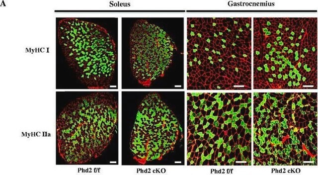
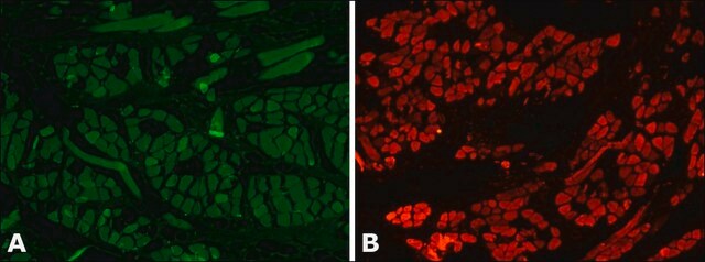
Immunofluorescence Analysis: A representative lot immunostained type 2A fibers in mouse hindlimb muscle cryosections encompassing soleus and surrounding tissues (Kurapati, R., et al. (2012). Hum. Mol. Genet. 21(8):1706-1724).

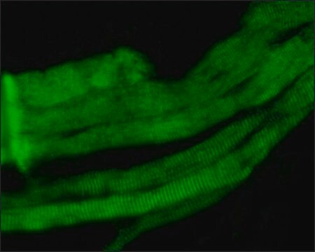
Immunofluorescence Analysis: A representative lot immunostained type 2A fibers in rat soleus muscle cryosections following Bupivacaine-induced muscle regeneration. In tetrodotoxin/TTX-paralyzed-regenerated muscles type 2A MHC was not expressed (Midrio, M., et al. (2002). Basic Appl. Myol. 12(2): 77-80).

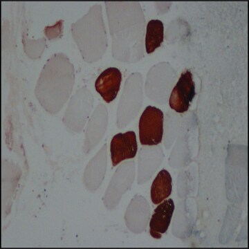
Immunohistochemistry Analysis: A representative lot detected type IIA myosin heavy chain (MyHC) in human masseter (jaw) muscle cryosections (Horton, M.J., et al. (2001). Arch. Oral Biol. 46(11):1039-1050).

Immunohistochemistry Analysis: Representative lots immunostained type 2A, but not type 1, 2B, or 2X, fibers in soleus (rat) and tibialis (mouse, rat, and Mgray short-tailed opossum/Monodelphis domestica) anterior muscle cryosections. Clone SC-71 failed to stain guinea pig tibialis sections (Sciote, J.J., and Rowlerson, A. (1998). Anat. Rec. 251(4):548-562; Gorza, L. (1990). J. Histochem Cytochem. 38(2):257-265; Schiaffino, S., et al. (1989). J. Muscle Res. Cell Motil. 10(3):197-205).

Western Blotting Analysis: A representative lot detected type IIA myosin heavy chain (MyHC) in human masseter (jaw) single muscle fibres extract (Horton, M.J., et al. (2001). Arch. Oral Biol. 46(11):1039-1050).

Western Blotting Analysis: A representative lot detected myosin heavy chain (MHC) in myosin preparations from rat diaphragm, as well as the light meromyosin and rod, but not heavy meromyosin or S-1, fragments of MHC (Schiaffino, S., et al. (1989). J. Muscle Res. Cell Motil. 10(3):197-205).

Quality

Identity Confirmation by Isotyping Test.

Isotyping Analysis: The identity of this monoclonal antibody is confirmed by isotyping test to be mouse IgG1 .

Target description

~225 kDa observed. 223.3 kDa (bovine), 223.0 (human), 223.2 kDa (mouse) calculated. Uncharacterized bands may be observed in some lysate(s).

Physical form

Format: Purified
Protein G purified.
Purified mouse IgG1 in buffer containing 0.1 M Tris-Glycine (pH 7.4), 150 mM NaCl with 0.05% sodium azide.

Storage and Stability

Stable for 1 year at 2-8°C from date of receipt.

Other Notes

Concentration: Please refer to lot specific datasheet.

Disclaimer

Unless otherwise stated in our catalog or other company documentation accompanying the product(s), our products are intended for research use only and are not to be used for any other purpose, which includes but is not limited to, unauthorized commercial uses, in vitro diagnostic uses, ex vivo or in vivo therapeutic uses or any type of consumption or application to humans or animals.

¿No encuentra el producto adecuado?  

Pruebe nuestro Herramienta de selección de productos.

Storage Class

12 - Non Combustible Liquids

wgk_germany

WGK 1


Certificados de análisis (COA)

Busque Certificados de análisis (COA) introduciendo el número de lote del producto. Los números de lote se encuentran en la etiqueta del producto después de las palabras «Lot» o «Batch»

¿Ya tiene este producto?

Encuentre la documentación para los productos que ha comprado recientemente en la Biblioteca de documentos.

Visite la Librería de documentos

Youngmok C Jang et al.
FASEB journal : official publication of the Federation of American Societies for Experimental Biology, 24(5), 1376-1390 (2009-12-31)
Oxidative stress has been implicated in the etiology of age-related muscle loss (sarcopenia). However, the underlying mechanisms by which oxidative stress contributes to sarcopenia have not been thoroughly investigated. To directly examine the role of chronic oxidative stress in vivo
Tomoya Uchimura et al.
Cell reports. Medicine, 2(6), 100298-100298 (2021-07-02)
Duchenne muscular dystrophy (DMD) is a muscle degenerating disease caused by dystrophin deficiency, for which therapeutic options are limited. To facilitate drug development, it is desirable to develop in vitro disease models that enable the evaluation of DMD declines in contractile

Nuestro equipo de científicos tiene experiencia en todas las áreas de investigación: Ciencias de la vida, Ciencia de los materiales, Síntesis química, Cromatografía, Analítica y muchas otras.

Póngase en contacto con el Servicio técnico