General description
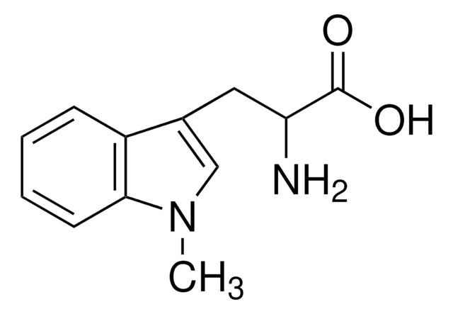
Indoleamine 2,3-dioxygenase 2 (UniProt: Q8R0V5; also known as IDO-2, Indoleamine 2,3-dioxygenase-like protein 1, Indoleamine-pyrrole 2,3-dioxygenase-like protein 1) is encoded by the Ido2 (also known as Indol1) gene (Gene ID: 209176) in murine species. IDO-2 is expressed mainly in antigen-presenting immune cells, liver, kidney, brain, and placenta. It is highly expressed in kidney, followed by epididymis and liver and is also detected in the tails of the spermatozoa. It catalyzes the first and rate-limiting step in the kynurenine pathway of tryptophan catabolism. IDO-1 and IDO-2 are 2 distinct enzymes which catalyze the same reaction. However, the Km for IDO-2 for tryptophan is much higher than that of IDO-1. IDO-2 may play a role as a negative regulator of IDO-1 by competing for heme-binding with IDO-1. IDO-2 activity is inhibited by 1MT (1-methyl-tryptophan) and MTH-trp (methylthiohydantoin-DL-tryptophan). IDO-2 is also involved in immune regulation. IDO-2 knockout mice do not show any apparent defects in their embryonic development or hematopoietic differentiation and have wild-type profiles for kynurenine in blood serum and for immune cells in spleen, lymph nodes, peritoneum, thymus and bone marrow. However, the knockout mice exhibit defects in IDO-mediated T-cell regulation and inflammatory responses. (Ref.: Mbongue JC, et al. (2015). Vaccines (Basel). 3(3):703-29).
Specificity
Clone 4-3 detects IDO-2 in mouse. It targets an epitope within 21 amino acids in the C-terminal region.
Immunogen
KLH-conjugated linear peptide/recombinant protein corresponding to 21 amino acids from the C-terminal region of murine Indoleamine 2,3-dioxygenase 2 (IDO-2).
Application
Anti-IDO-2, clone 4-3, Cat. No. MABF2055, is a mouse monoclonal antibody that detects IDO2 and has been tested for use in Western Blotting.
Research Category
Inflammation & Immunology
Western Blotting Analysis: 0.5 µg/mL from a represesntative lot detected IDO-2 in 10 µg of mouse kidney tissue lysate.
Quality
Evaluated by Western Blotting in mouse liver tissue lysate.
Western Blotting Analysis: 0.5 µg/mL of this antibody detected IDO-2 in 10 µg of mouse liver tissue lysate.
Target description
~55 kDa observed; 45.25 kDa calculated. Uncharacterized bands may be observed in some lysate(s).
Physical form
Format: Purified
Protein G purified
Purified mouse monoclonal antibody IgG1 in buffer containing 0.1 M Tris-Glycine (pH 7.4), 150 mM NaCl with 0.05% sodium azide.
Storage and Stability
Stable for 1 year at 2-8°C from date of receipt.
Other Notes
Concentration: Please refer to lot specific datasheet.
Disclaimer
Unless otherwise stated in our catalog or other company documentation accompanying the product(s), our products are intended for research use only and are not to be used for any other purpose, which includes but is not limited to, unauthorized commercial uses, in vitro diagnostic uses, ex vivo or in vivo therapeutic uses or any type of consumption or application to humans or animals.