Skip to Content
Merck
All Photos(4)

Key Documents

SAB4200753

Sigma-Aldrich

Anti-phospho-AKT (pThr450) antibody, Mouse monoclonal

clone AK-11, purified from hybridoma cell culture

Synonym(s):

Anti-Protein Kinase B, Anti-V-Akt Murine Thymoma Viral Oncogene Homolog 1

Sign Into View Organizational & Contract Pricing


About This Item

UNSPSC Code:
12352203
NACRES:
NA.44
clone:
AK-11, monoclonal
application:
IF
IP
species reactivity:
mouse, chicken, rat, human
technique(s):
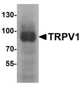
immunoblotting: 1-2 μg/mL using human breast cancer cell line MCF-7 extract
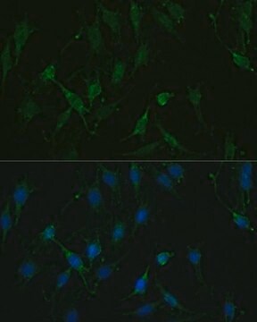
immunofluorescence: 1-2 μg/mL using mouse embryo fibroblast NIH-3T3 cells
immunoprecipitation (IP): 2-4 μg/test using whole extract of mouse embryo fibroblast NIH-3T3 cell line
citations:
4

biological source

mouse

Quality Level

antibody form

purified from hybridoma cell culture

antibody product type

primary antibodies

clone

AK-11, monoclonal

form

buffered aqueous solution

mol wt

~56 kDa

species reactivity

mouse, chicken, rat, human

concentration

~1.0 mg/mL

technique(s)

immunoblotting: 1-2 μg/mL using human breast cancer cell line MCF-7 extract
immunofluorescence: 1-2 μg/mL using mouse embryo fibroblast NIH-3T3 cells
immunoprecipitation (IP): 2-4 μg/test using whole extract of mouse embryo fibroblast NIH-3T3 cell line

isotype

IgG2a

UniProt accession no.

shipped in

dry ice

storage temp.

−20°C

target post-translational modification

phosphorylation (pThr450)

Gene Information

human ... AKT1(207)

General description

Anti-phospho-AKT (pThr450) antibody, mouse monoclonal (mouse IgG2a isotype) is derived from the AK-11 hybridoma produced by the fusion of mouse myeloma cells and splenocytes from an immunized BALB/C mouse. Serine/threonine kinase (AKT) also known as protein kinase B (PKB) belongs to the family of serine/threonine kinases. It consists of three isoforms such as AKT1, AKT2 and AKT3. AKT1 gene is located on the human chromosome at 14q32.33.

Specificity

Anti-phospho-AKT (pThr450) antibody, Mouse monoclonal specifically recognizes all three AKT isoforms phosphorylated at Thr450 (AKT1, AKT2 and AKT3).

Immunogen

Synthetic peptide corresponding to human AKT1 pThr450

Application

Anti-phospho-AKT (pThr450) antibody, mouse monoclonal may be used in:
  • immunoblotting
  • immunofluorescence
  • immunoprecipitation

Biochem/physiol Actions

Serine/threonine kinase (AKT) plays an important role in the control of cell cycle and cell proliferation. It is also involved in cell differentiation and apoptosis signaling pathways. AKT is rapidly activated in response to cell stimulation by several growth factors, such as insulin, peroxyvanadate or cellular stresses (such as heat shock). Following activation, the phosphorylated AKT is relocated to the nucleus. Lack of phosphorylation at Thr450 enhances AKT ubiquitination and degradation.

Physical form

Solution in 0.01 M phosphate buffered saline, pH 7.4, containing 15 mM sodium azide.

Storage and Stability

For continuous use, store at 2–8 °C for up to one month. For extended storage, freeze in working aliquots. Repeated freezing and thawing is not recommended. If slight turbidity occurs upon prolonged storage, clarify the solution by centrifugation before use. Working dilution samples should be discarded if not used within 12 hours.

Disclaimer

Unless otherwise stated in our catalog, our products are intended for research use only and are not to be used for any other purpose, which includes but is not limited to, unauthorized commercial uses, in vitro diagnostic uses, ex vivo or in vivo therapeutic uses or any type of consumption or application to humans or animals.

Not finding the right product?  

Try our Product Selector Tool.

Storage Class Code

10 - Combustible liquids

WGK

WGK 2

Flash Point(F)

Not applicable

Flash Point(C)

Not applicable


Choose from one of the most recent versions:

Certificates of Analysis (COA)

Lot/Batch Number

Don't see the Right Version?

If you require a particular version, you can look up a specific certificate by the Lot or Batch number.

Already Own This Product?

Find documentation for the products that you have recently purchased in the Document Library.

Visit the Document Library

H Dudek et al.
Science (New York, N.Y.), 275(5300), 661-665 (1997-01-31)
A signaling pathway was delineated by which insulin-like growth factor 1 (IGF-1) promotes the survival of cerebellar neurons. IGF-1 activation of phosphoinositide 3-kinase (PI3-K) triggered the activation of two protein kinases, the serine-threonine kinase Akt and the p70 ribosomal protein
Zhi-Ping Yang et al.
World journal of gastroenterology, 21(36), 10385-10399 (2015-10-01)
To investigate the anti-tumor effects of equol in gastric cancer cells and the underlying molecular mechanisms. MGC-803 cells were employed for in vitro experiments in this study. Cells were treated with control (vehicle, 0.1% DMSO) or equol under specified dose
Won Jun Oh et al.
The EMBO journal, 29(23), 3939-3951 (2010-11-04)
The mechanisms that couple translation and protein processing are poorly understood in higher eukaryotes. Although mammalian target of rapamycin (mTOR) complex 1 (mTORC1) controls translation initiation, the function of mTORC2 in protein synthesis remains to be defined. In this study
A Khwaja et al.
The EMBO journal, 16(10), 2783-2793 (1997-05-15)
Upon detachment from the extracellular matrix, epithelial cells enter into programmed cell death, a phenomenon known as anoikis, ensuring that they are unable to survive in an inappropriate location. Activated ras oncogenes protect cells from this form of apoptosis. The
L Xiao et al.
Cell death and differentiation, 17(9), 1448-1462 (2010-02-27)
AKT pathway has a critical role in mediating signaling transductions for cell proliferation, differentiation and survival. Previous studies have shown that AKT activation is achieved through a series of phosphorylation steps: first, AKT is phosphorylated at Thr-450 by JNK kinases

Our team of scientists has experience in all areas of research including Life Science, Material Science, Chemical Synthesis, Chromatography, Analytical and many others.

Contact Technical Service