MIDI67
PKH67 Green Fluorescent Cell Linker Midi Kit for General Cell Membrane Labeling
Distributed for Phanos Technologies
Synonym(s):
Green PKH membrane midi labeling kit
About This Item
Recommended Products
packaging
pkg of 1 kit
Quality Level
manufacturer/tradename
Distributed for Phanos Technologies
storage condition
protect from light
technique(s)
flow cytometry: suitable
fluorescence
λex 490 nm; λem 502 nm (PKH67 dye)
application(s)
cell analysis
detection
detection method
fluorometric
shipped in
ambient
storage temp.
room temp
General description
Slow loss of fluorescence has been observed in in vivo studies using PKH1 and PKH2. PKH67 may exhibit similar properties since this behavior appears to be characteristic of green cell linker dyes, but not red cell linker dyes. Correlation of in vitro cell membrane retention with in vivo fluorescence half life in non-dividing cells predicts an in vivo fluorescence half life of 10-12 days for PKH67. Other green cell linker dyes with similar half lives have been used to monitor in vivo lymphocyte and macrophage trafficking over periods of 1-2 months, suggesting that PKH67 will also be useful for in vivo tracking studies of moderate length.
Application
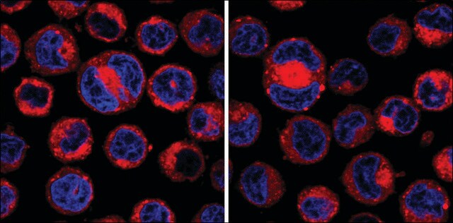
- labeling exosomes
- extracellular vesicles (EVs)l
- normal and sickle red blood cells (RBCs)
- Raji cells
Linkage
Legal Information
Kit Components Only
- PKH67 Dye 2 x 0.1
- Diluent C 6 x 10
Signal Word
Danger
Hazard Statements
Precautionary Statements
Hazard Classifications
Eye Irrit. 2 - Flam. Liq. 2
Storage Class Code
3 - Flammable liquids
Flash Point(F)
57.2 °F - closed cup
Flash Point(C)
14 °C - closed cup
Choose from one of the most recent versions:
Already Own This Product?
Find documentation for the products that you have recently purchased in the Document Library.
Customers Also Viewed
Articles
Optimal staining is a key component for studying tumorigenesis and progression. Learn useful tips and techniques for dye applications, including examples from recent studies.
A video about how you can use fluorescent cell tracking dyes in combination with flow and image cytometry to study interactions and fates of different cell types in vitro and in vivo.
PKH and CellVue® Fluorescent Cell Linker Kits provide fluorescent labeling of live cells over an extended period of time, with no apparent toxic effects.
Our team of scientists has experience in all areas of research including Life Science, Material Science, Chemical Synthesis, Chromatography, Analytical and many others.
Contact Technical Service