Skip to Content
Merck
All Photos(1)

Key Documents

MABT827

Sigma-Aldrich

Anti-Dystrophin Antibody, clone 2C6 (MANDYS106)

clone 2C6 (MANDYS106), from mouse

Synonym(s):

Dystrophin

Sign Into View Organizational & Contract Pricing


About This Item

UNSPSC Code:
12352203
eCl@ss:
32160702
NACRES:
NA.41

biological source

mouse

Quality Level

antibody form

purified antibody

antibody product type

primary antibodies

clone

2C6 (MANDYS106), monoclonal

species reactivity

human

technique(s)

immunofluorescence: suitable
immunohistochemistry: suitable
western blot: suitable

isotype

IgG2aκ

NCBI accession no.

UniProt accession no.

shipped in

wet ice

target post-translational modification

unmodified

Gene Information

human ... DMD(1756)

General description

Dystrophin (UniProt P11532) is encoded by the DMD (also known as BMD, CMD3B, DXS142, DXS164, DXS206, DXS230, DXS239, DXS268, DXS269, DXS270, DXS272, MRX85) gene (Gene ID 1756) in human. Dystrophin is localized to the inner part of the muscle fiber cell membrane (sarcolemma) and plays an important role in stabilizing the muscle fiber against the mechanical forces of muscle contraction by providing a shock-absorbing connection between the cytoskeleton and the extracellular matrix. Duchenne muscular dystrophy (DMD) is caused by gene mutations that disrupt the open reading frame (ORF) and prevent the full translation of dystrophin. ORF restoration by exon skipping using antisense oligonucleotides is designed to transform the DMD phenotype to that of the milder disorder, Becker muscular dystrophy (BMD), which is typically caused by in-frame dystrophin deletions that allow the production of an internally deleted, but partially functional dystrophin.

Specificity

Detects dystrophin spliced isoforms 1-4, but not isoforms 5-10, or utrophin. Positive muscle membrane staining of tissue samples from healthy donors, reduced staining of Becker muscular dystrophy (BMD) biopsies, and no staining is seen with Duchenne muscular dystrophy (DMD) biopsy samples.

Immunogen

Epitope: Exon 43-coded pectrin-like repeat 16 region
TrpE-tagged recombinant protein corresponding to the Exon 43-coded pectrin-like repeat 16 region of human Dystrophin.

Application

Anti-Dystrophin Antibody, clone 2C6 (MANDYS106) is an antibody against Dystrophin for use in Immunohistochemistry, Immunofluorescence, Western Blotting.
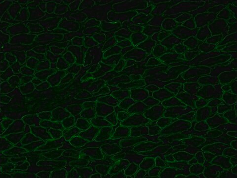
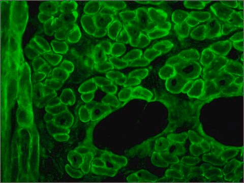
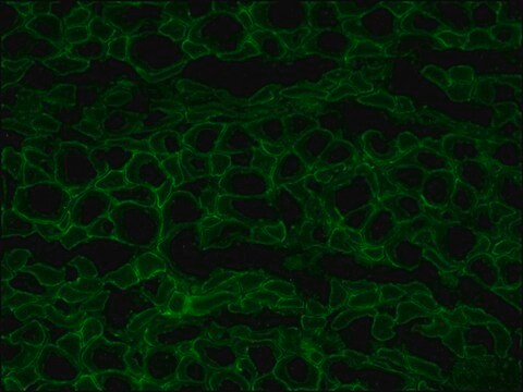
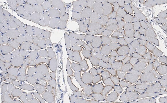
Immunohistochemistry Analysis: A represenative lot stained sarcolemma of muscle fiber cells in tissue samples from healthy donors, while much reduced staining was observed in biopsy samples from patients with Becker muscular dystrophy (BMD) and no staining is seen with Duchenne muscular dystrophy (DMD) biopsy samples (Anthony, K., et al. (2011). Brain. 134(Pt 12):3547-3559).
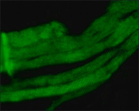
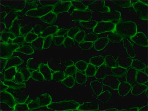
Immunofluorescence Analysis: A represenative lot was employed together with a spectrin antibody in dual immunofluorescent sarcolemma staining for assessing dystrophin levels of muscle fiber cells in muscle biopsies from healthy donors and Becker muscular dystrophy (BMD) patients (Beekman, C., et al. (2014). PLoS One. 9(9):e107494).
Research Category
Cell Structure
Research Sub Category
Adhesion (CAMs)

Quality

Evaluated by Immunohistochemistry in human skeletal muscle myocytes.

Immunohistochemistry Analysis: A 1:50 dilution of this antibody detected Dystrophin in human skeletal muscle myocytes.

Target description

~427 kDa observed

Physical form

Format: Purified
Protein G Purified
Purified mouse monoclonal IgG2aκ antibody in buffer containing 0.1 M Tris-Glycine (pH 7.4), 150 mM NaCl with 0.05% sodium azide.

Storage and Stability

Stable for 1 year at 2-8°C from date of receipt.

Other Notes

Concentration: Please refer to lot specific datasheet.

Disclaimer

Unless otherwise stated in our catalog or other company documentation accompanying the product(s), our products are intended for research use only and are not to be used for any other purpose, which includes but is not limited to, unauthorized commercial uses, in vitro diagnostic uses, ex vivo or in vivo therapeutic uses or any type of consumption or application to humans or animals.

Not finding the right product?  

Try our Product Selector Tool.

Storage Class Code

12 - Non Combustible Liquids

WGK

WGK 1

Flash Point(F)

Not applicable

Flash Point(C)

Not applicable


Certificates of Analysis (COA)

Search for Certificates of Analysis (COA) by entering the products Lot/Batch Number. Lot and Batch Numbers can be found on a product’s label following the words ‘Lot’ or ‘Batch’.

Already Own This Product?

Find documentation for the products that you have recently purchased in the Document Library.

Visit the Document Library

Menglong Chen et al.
Genome medicine, 13(1), 57-57 (2021-04-14)
Mutations in the DMD gene encoding dystrophin-a critical structural element in muscle cells-cause Duchenne muscular dystrophy (DMD), which is the most common fatal genetic disease. Clustered regularly interspaced short palindromic repeat (CRISPR)-mediated gene editing is a promising strategy for permanently
Diane E Frank et al.
Neurology, 94(21), e2270-e2282 (2020-03-07)
To report safety, pharmacokinetics, exon 53 skipping, and dystrophin expression in golodirsen-treated patients with Duchenne muscular dystrophy (DMD) amenable to exon 53 skipping. Part 1 was a randomized, double-blind, placebo-controlled, 12-week dose titration of once-weekly golodirsen; part 2 is an
Dystrophin quantification and clinical correlations in Becker muscular dystrophy: implications for clinical trials.
Anthony, K; Cirak, S; Torelli, S; Tasca, G; Feng, L; Arechavala-Gomeza, V; Armaroli et al.
Brain null
Valentina Sardone et al.
PloS one, 13(3), e0194540-e0194540 (2018-03-27)
Clinical trials using strategies aimed at inducing dystrophin expression in Duchenne muscular dystrophy (DMD) are underway or at advanced planning stage, including splice switching antisense oligonucleotides (AON), drugs to induce read-through of nonsense mutations and viral mediated gene therapy. In
Chantal Beekman et al.
PloS one, 9(9), e107494-e107494 (2014-09-23)
Duchenne muscular dystrophy (DMD) is characterized by the absence or reduced levels of dystrophin expression on the inner surface of the sarcolemmal membrane of muscle fibers. Clinical development of therapeutic approaches aiming to increase dystrophin levels requires sensitive and reproducible

Our team of scientists has experience in all areas of research including Life Science, Material Science, Chemical Synthesis, Chromatography, Analytical and many others.

Contact Technical Service