Skip to Content
Merck
All Photos(1)

Key Documents

MABN852

Sigma-Aldrich

Anti-Amyloid Precursor Protein (APP) Antibody

mouse monoclonal, 2B3

Synonym(s):

Amyloid-beta A4 protein, ABPP, APPI, Alzheimer disease amyloid protein, Amyloid precursor protein, Amyloid-beta precursor protein, Cerebral vascular amyloid peptide, CVAP, PreA4, Protease nexin-II, PN-II

Sign Into View Organizational & Contract Pricing


About This Item

UNSPSC Code:
12352203
eCl@ss:
32160702
NACRES:
NA.43
clone:
2B3, monoclonal
application:
ELISA
ICC
WB
inhibition assay
species reactivity:
mouse, human
technique(s):
ELISA: suitable
immunocytochemistry: suitable
inhibition assay: suitable
western blot: suitable
citations:

Product Name

Anti-APP Antibody, clone 2B3, clone 2B3, from mouse

biological source

mouse

antibody form

purified immunoglobulin

antibody product type

primary antibodies

clone

2B3, monoclonal

species reactivity

mouse, human

species reactivity (predicted by homology)

rat (based on 100% sequence homology)

packaging

antibody small pack of 25 μg

technique(s)

ELISA: suitable
immunocytochemistry: suitable
inhibition assay: suitable
western blot: suitable

isotype

IgG1κ

NCBI accession no.

UniProt accession no.

target post-translational modification

unmodified

Gene Information

human ... APP(351)

General description

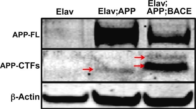
Amyloid-beta A4 protein (UniProt: P05067; also known as ABPP, APPI, APP, Alzheimer disease amyloid protein, Amyloid precursor protein, Amyloid-beta precursor protein, Cerebral vascular amyloid peptide, CVAP, PreA4, Protease nexin-II, PN-II) is encoded by the APP (also known as A4, AD1) gene (Gene ID: 351) in human. APP undergoes extensive post-translational modification including glycosylation, phosphorylation, and tyrosine sulfation, as well as many types of proteolytic processing to generate peptide fragments. APP is proteolytically processed under normal cellular conditions by alpha-secretase or beta-secretase to generate and release soluble APP peptides, S-APP-alpha and S-APP-beta, and the retention of corresponding membrane-anchored C-terminal fragments, C80, C83 and C99. Subsequent processing of C80 and C83 by gamma-secretase yields P3 peptides. In Alzheimer s disease processing of C99 generates amyloid-beta 40 (Abeta40) and amyloid-beta 42 (Abeta42) that form amyloid plaques. Beta-amyloid peptides are lipophilic metal chelators with metal-reducing activity. They bind transient metals such as copper, zinc and iron. APP can also be cleaved by caspases during neuronal apoptosis. Cleavage at Asp-739 by either caspase-6, -8 or -9 results in the production of the neurotoxic C31 peptide and the increased production of beta-amyloid peptides. In addition to its obvious role in Alzheimer′s disease, the most-substantiated role for APP is in synaptic formation and repair. Its expression is upregulated during neuronal differentiation and after neural injury.

Specificity

Clone 2B3 specifically detects Amyloid-beta A4 protein in human and murine neurons. It targets an epitope within 15 amino acids including the beta-secretase cleavage site.

Immunogen

A linear peptide corresponding to 15 amino acids from the C-terminal region of human Amyloid-beta A4 protein.
Epitope: extracellular domain

Application

Anti-APP, clone 2B3, Cat. No. MABN852, is a highly specific mouse monoclonal antibody that targets Amyloid beta A4 protein and has been tested for use in ELISA, Immunocytochemistry, Inhibiton Assay, and Western Blotting.
ELISA Analysis: A representative lot detected APP in ELISA applications (Thomas, R.S., et. al. (2011). FEBS J. 278(1):167-78; Thomas, R.S., et. al. (2013). Neuroreport. 24(18):1058-61).

Inhibition Analysis: A representative lot inhibited beta amyloid production in MOG-G-UVW culture media (Thomas, R.S., et. al. (2011). FEBS J. 278(1):167-78).

Western Blotting Analysis: A representative lot detected APP in Western Blotting applications (Thomas, R.S., et. al. (2011). FEBS J. 278(1):167-78).

Immunocytochemistry Analysis: A representative lot detected APP in Immunocytochemistry applications (Thomas, R.S., et. al. (2013). Neuroreport. 24(18):1058-61).
Research Category
Neuroscience

Quality

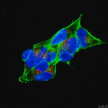
Evaluated by Immunocytochemistry in mouse cortical neurons.

Immunocytochemistry Analysis: A 1:100 dilution of this antibody detected APP in mouse cortical neurons.

Target description

86.94 kDa calculated.

Physical form

Format: Purified
Protein G purified
Purified mouse monoclonal antibody IgG1 in PBS without preservatives.

Storage and Stability

Stable for 1 year at -20°C from date of receipt. Handling Recommendations: Upon receipt and prior to removing the cap, centrifuge the vial and gently mix the solution. Aliquot into microcentrifuge tubes and store at -20°C. Avoid repeated freeze/thaw cycles, which may damage IgG and affect product performance.

Other Notes

Concentration: Please refer to lot specific datasheet.

Disclaimer

Unless otherwise stated in our catalog or other company documentation accompanying the product(s), our products are intended for research use only and are not to be used for any other purpose, which includes but is not limited to, unauthorized commercial uses, in vitro diagnostic uses, ex vivo or in vivo therapeutic uses or any type of consumption or application to humans or animals.

Not finding the right product?  

Try our Product Selector Tool.


Certificates of Analysis (COA)

Search for Certificates of Analysis (COA) by entering the products Lot/Batch Number. Lot and Batch Numbers can be found on a product’s label following the words ‘Lot’ or ‘Batch’.

Already Own This Product?

Find documentation for the products that you have recently purchased in the Document Library.

Visit the Document Library

Our team of scientists has experience in all areas of research including Life Science, Material Science, Chemical Synthesis, Chromatography, Analytical and many others.

Contact Technical Service