Skip to Content
Merck
All Photos(3)

Key Documents

C7805

Sigma-Aldrich

Monoclonal Anti-Collagen, Type III antibody produced in mouse

clone FH-7A, ascites fluid

Synonym(s):

Anti-Collagen III antibody

Sign Into View Organizational & Contract Pricing


About This Item

MDL number:
UNSPSC Code:
12352203
NACRES:
NA.41

biological source

mouse

Quality Level

conjugate

unconjugated

antibody form

ascites fluid

antibody product type

primary antibodies

clone

FH-7A, monoclonal

mol wt

antigen 70 kDa

contains

15 mM sodium azide

species reactivity

human, rat

technique(s)

dot blot: suitable
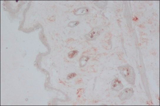
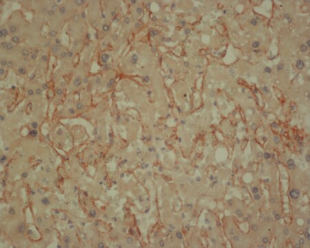
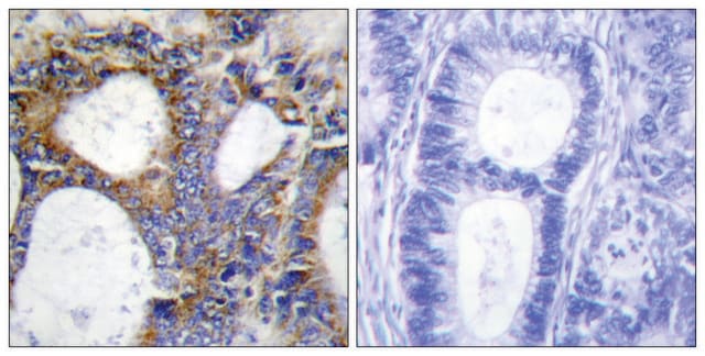
immunohistochemistry (frozen sections): 1-4,000 using frozen sections of human skin
indirect ELISA: suitable
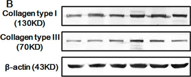
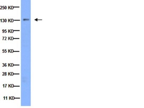
western blot: suitable using a denatured-reduced preparation

isotype

IgG1

UniProt accession no.

shipped in

dry ice

storage temp.

−20°C

target post-translational modification

unmodified

Gene Information

human ... COL3A1(1281)
rat ... Col3a1(84032)

General description

Collagen is a fibrous protein present in the extracellular framework of all vertebrates. Collagen type III is a 300 kD molecule that belongs to fibrillar collagens family found mainly in skin, blood vessels, liver, placenta, tongue and thymus. This collagen is directly or indirectly involved in several genetic diseases like Ehlers-Danlos type IV disease. Monoclonal anti-collagen type III antibody can be used to determine the interstitial collagen I and III content in total protein homogenates. It is also used to study the immunolocalization of collagen type III in normal and neoplastic tissue. Mouse monoclonal anti-collagen type III antibody reacts specifically with native and denatured collagen type III but not with collagen types I, II, IV, V, VI, and X.
Collagen is a fibrous protein present in the extracellular framework of all vertebrates. Collagen type III is a 300 kD molecule that belongs to fibrillar collagens family found mainly in skin, blood vessels, liver, placenta, tongue and thymus. This collagen is directly or indirectly involved in several genetic diseases like Ehlers-Danlos type IV disease. Mouse monoclonal anti-collagen type III antibody reacts specifically with native and denatured collagen type III but not with collagen types I, II, IV, V, VI, and X. This product has also shown reactivity for rat and human collagen type III.
Monoclonal Anti-Collagen Type III (mouse IgG1 isotype) is derived from the FH-7A hybridoma produced by the fusion of mouse myeloma cells and splenocytes from BALB/c mice immunized with human collagen type III.

Specificity

Monoclonal Anti-Collagen Type III reacts specifically with native and denatured collagen type III. It does not recognize collagen types I, II, IV, V, VI, and X.

Immunogen

human collagen type III

Application

Monoclonal Anti-Collagen, Type III antibody produced in mouse has also been used in enzyme linked immunosorbent assay (ELISA) and immunohistochemistry.
Monoclonal anti-collagen type III antibody has been used in immunofluorescence and western blot.

Biochem/physiol Actions

This collagen is directly or indirectly involved in several genetic diseases like Ehlers-Danlos type IV disease. Monoclonal anti-collagen type III antibody can be used to determine the interstitial collagen I and III content in total protein homogenates.

Disclaimer

Unless otherwise stated in our catalog or other company documentation accompanying the product(s), our products are intended for research use only and are not to be used for any other purpose, which includes but is not limited to, unauthorized commercial uses, in vitro diagnostic uses, ex vivo or in vivo therapeutic uses or any type of consumption or application to humans or animals.

Not finding the right product?  

Try our Product Selector Tool.

Storage Class Code

10 - Combustible liquids

WGK

WGK 2

Flash Point(F)

Not applicable

Flash Point(C)

Not applicable


Certificates of Analysis (COA)

Search for Certificates of Analysis (COA) by entering the products Lot/Batch Number. Lot and Batch Numbers can be found on a product’s label following the words ‘Lot’ or ‘Batch’.

Already Own This Product?

Find documentation for the products that you have recently purchased in the Document Library.

Visit the Document Library

The use of mesenchymal stem cells in collagen-based scaffolds for tissue-engineered repair of tendons
Butler DL, et al.
Nature Protocols, 5(5), 849-849 (2010)
Corinne N Riggin et al.
Journal of orthopaedic research : official publication of the Orthopaedic Research Society, 37(9), 2035-2042 (2019-05-02)
Overuse-induced tendinopathy is highly prevalent in the general population. Percutaneous fenestration, or dry needling, techniques have been increasing in popularity, but despite their current use, there are no controlled laboratory studies to provide fundamental support for this practice. The objective
Ana I Gonçalves et al.
PloS one, 8(12), e83734-e83734 (2014-01-05)
Current treatments for tendon injuries often fail to fully restore joint biomechanics leading to the recurrence of symptoms, and thus resulting in a significant health problem with a relevant social impact worldwide. Cell-based approaches involving the use of stem cells
Advanced glycation end products as a source of artifacts in immunoenzymatic methods
Kuzan A, et al.
Glycoconjugate Journal, 35(1), 95-103 (2018)
Fluorescently labeled collagen binding proteins allow specific visualization of collagen in tissues and live cell culture
Krahn KN, et al.
Analytical Biochemistry, 350(2), 177-185 (2006)

Our team of scientists has experience in all areas of research including Life Science, Material Science, Chemical Synthesis, Chromatography, Analytical and many others.

Contact Technical Service