Skip to Content
Merck
All Photos(1)

Documents

05-521

Sigma-Aldrich

Anti-TRF2 Antibody, clone 4A794

clone 4A794, Upstate®, from mouse

Synonym(s):

Anti-TRBF2, Anti-TRF2

Sign Into View Organizational & Contract Pricing


About This Item

UNSPSC Code:
12352203
eCl@ss:
32160702
NACRES:
NA.41

biological source

mouse

Quality Level

antibody form

purified immunoglobulin

antibody product type

primary antibodies

clone

4A794, monoclonal

species reactivity

rat, human

manufacturer/tradename

Upstate®

technique(s)

immunocytochemistry: suitable
western blot: suitable

isotype

IgG1

NCBI accession no.

UniProt accession no.

shipped in

dry ice

target post-translational modification

unmodified

Gene Information

human ... TERF2(7014)

General description

TRF2 is a ubiquitously expressed protein that is implicated in the control of telomere length. TRF2, like TRF1, contains a Myb-related DNA binding motif. It binds to duplex TTAGGG repeats and is localised to all human telomeres in metaphase chromosomes. TRF2 is thought to protect chromosome ends by maintaining the correct structure at telomere termini. The use of mutant forms of TRF2 has implicated a role for TRF2 in the prevention of senescence in primary human cells. Recently, it has been shown that inhibition of TRF2 resulted in apoptosis in a subset of mammalian cell types. TRF2 is overexpressed in a number of human tumors and overexpression of TRF2 has been shown to be oncogenic in a mouse model of carcinogenesis.

Specificity

Recognizes TRF2.

Immunogen

His-tagged, fusion-protein, corresponding to full-length TRF2 (Telomeric Repeat binding Factor 2), expressed in baculovirus

Application

Anti-TRF2 Antibody, clone 4A794 is a Mouse Monoclonal Antibody for detection of TRF2 also known as Telomeric repeat-binding factor 1 & has been tested in ICC & WB.
Research Category
Epigenetics & Nuclear Function
Research Sub Category
Cell Cycle, DNA Replication & Repair

Quality

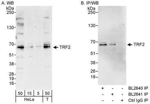
routinely evaluated by immunoblot on RIPA lysate of human Jurkat cells, HeLa nuclear extract, or rat L6 RIPA cell lysate

Target description

66 kDa

Physical form

Format: Purified
Protein G Purified
Protein G Purified mouse IgG1 in PBS with 0.05% sodium azide.

Storage and Stability

Maintain for 2 years at -20°C from date of shipment. Aliquot to avoid repeated freezing and thawing. For maximum recovery of product, centrifuge the original vial after thawing and prior to removing the cap.

Analysis Note

Control
Positive Antigen Control: Catalog #12-303, Jurkat cell lysate.

Legal Information

UPSTATE is a registered trademark of Merck KGaA, Darmstadt, Germany

Disclaimer

Unless otherwise stated in our catalog or other company documentation accompanying the product(s), our products are intended for research use only and are not to be used for any other purpose, which includes but is not limited to, unauthorized commercial uses, in vitro diagnostic uses, ex vivo or in vivo therapeutic uses or any type of consumption or application to humans or animals.

Not finding the right product?  

Try our Product Selector Tool.

Storage Class Code

12 - Non Combustible Liquids

WGK

WGK 2

Flash Point(F)

Not applicable

Flash Point(C)

Not applicable


Certificates of Analysis (COA)

Search for Certificates of Analysis (COA) by entering the products Lot/Batch Number. Lot and Batch Numbers can be found on a product’s label following the words ‘Lot’ or ‘Batch’.

Already Own This Product?

Find documentation for the products that you have recently purchased in the Document Library.

Visit the Document Library

S K Manna et al.
Journal of immunology (Baltimore, Md. : 1950), 165(9), 4927-4934 (2000-10-25)
We and others have reported that IFN-alpha potentiates the apoptotic effects of TNF through a mechanism that is not understood. Because the nuclear transcription factors NF-kappaB and AP-1 have recently been reported to mediate anti-apoptosis and cell survival, we hypothesized
Manale El Idrissi et al.
Journal of experimental & clinical cancer research : CR, 32, 64-64 (2013-09-12)
Among the numerous genetic defects associated with hepatocarcinogenesis, telomere abnormalities appear to play a role both in tumor promotion and maintenance. Telomeres, the chromosome extremities, are protected by specific proteins, the shelterin complex and by additional factors. Besides telomerase dysregulation
Clara Lopes Novo et al.
FASEB journal : official publication of the Federation of American Societies for Experimental Biology, 27(9), 3632-3642 (2013-06-05)
Telomeres are major regulators of genome stability and cell proliferation. A detailed understanding of the mechanisms involved in their maintenance is of foremost importance. Of those, telomere chromatin remodeling is probably the least studied; thus, we intended to explore the
Different telomere damage signaling pathways in human and mouse cells.
Agata Smogorzewska, Titia de Lange
The Embo Journal null
The cellular level of telomere dysfunction determines induction of senescence or apoptosis in vivo.
Andre Lechel, Ande Satyanarayana, Zhenyu Ju, Ruben R Plentz, Sonja Schaetzlein et al.
EMBO Reports null

Our team of scientists has experience in all areas of research including Life Science, Material Science, Chemical Synthesis, Chromatography, Analytical and many others.

Contact Technical Service