일반 설명
We are committed to bringing you greener alternative products, which adhere to one or more of The 12 Principles of Green Chemistry.This antibody is Preservative-free, produced without the harm or sacrifice of animals and exceptionally stable to allow for ambient shipping and storage if needed and thus aligns with "Waste Prevention", "Designing Safer Chemicals" and "Design for Energy Efficiency".
Click here for more information.
ZooMAb antibodies represent an entirely new generation of recombinant monoclonal antibodies.
Each ZooMAb antibody is manufactured using our proprietary recombinant expression system, purified to homogeneity, and precisely dispensed to produce robust and highly reproducible lot-to-lot consistency. Only top-performing clones are released for use by researchers. Each antibody is validated for high specificity and affinity across multiple applications, including its most commonly used application. ZooMAb antibodies are reliably available and ready to ship when you need them.
Learn more about ZooMAb here.특이성
Clone 13E6 is a ZooMAb rabbit recombinant monoclonal antibody that specifically detects Neuronal nuclei antigen. It targets an epitope with in the first 97 amino acids from the N-terminal region.
면역원
GST/His-tagged recombinant fragment corresponding to the first 97 amino acids from the N-terminal region of murine Neuronal nuclei antigen (NeuN).
애플리케이션
Anti-NeuN, clone 13E6, ZooMAb, Cat. No. ZRB377, is a highly specific recombinant Rabbit Monoclonal antibody that detects Neuronal nuclei antigen and has been tested for use in Immunocytochemistry, Immunofluorescence, Immunohistochemistry (Paraffin), and Western Blotting.
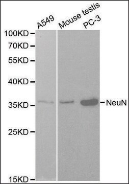
Western Blotting Analysis: A 1:1,000 dilution from a representative lot detected NeuN in Rat brain nuclear extract.
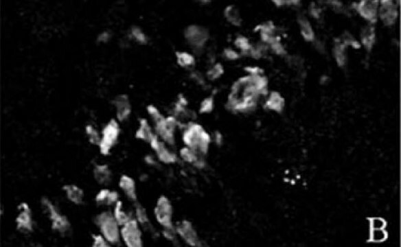
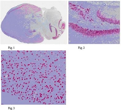
Immunofluorescence Analysis: A 1:100 dilution from a representative lot detected NeuN in mouse hippocampus FFPE and mouse cerebral cortex frozen sections.
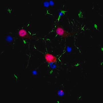
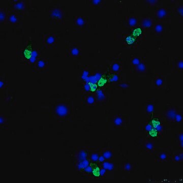
Immunocytochemistry Analysis: A 1:100 dilution from a representative lot detected NeuN in E18 rat cortical cells.
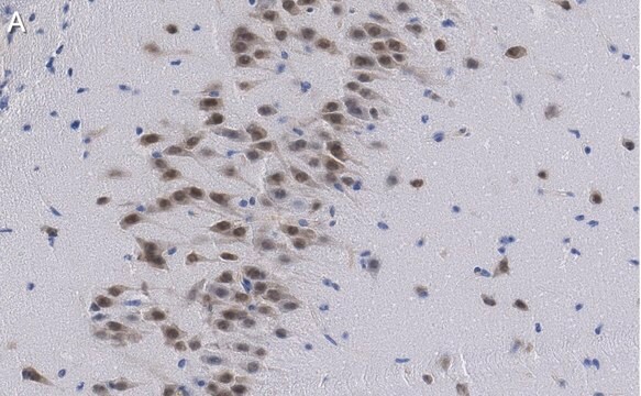
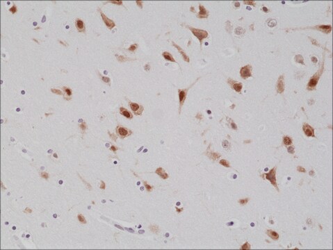
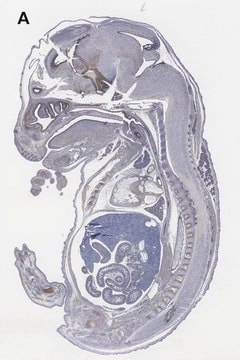
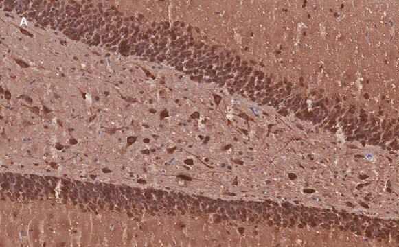
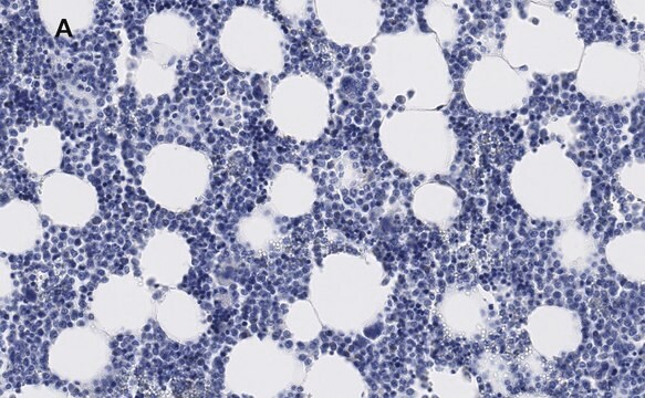
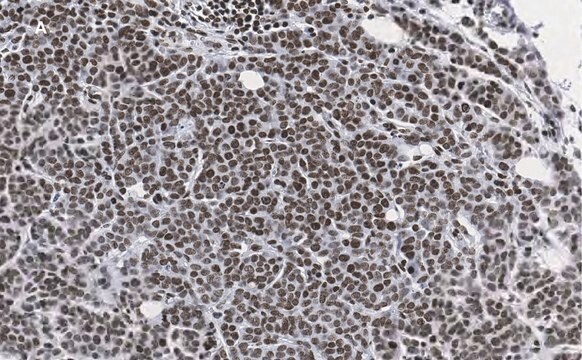
Immunohistochemistry (Paraffin) Analysis: A 1:100 dilution from a representative lot detected NeuN in mouse brain and human brain tissue sections.
표적 설명
RNA binding protein fox-1 homolog 3 (UniProt: Q8BIF2; also known as Fox-1 homolog C, Hexaribonucleotide-binding protein 3, Fox-3, Neuronal nuclei antigen, NeuN antigen) is encoded by the Rbfox3 (also known as D11Bwg0517e, Hrnbp3) gene (Gene ID: 52897) in murine species. The NeuN protein is localized in nuclei and perinuclear cytoplasm of most of the neurons in the central nervous system of mammals. It is widely expressed in brain, including in cerebral cortex, hippocampus, thalamus, caudate/putamen, cerebellum, as well as in the spinal cord. However, it is not detected in Cajal-Retzius cells in the neocortex, Purkinje cells, and photoreceptor cells. NeuN expression has been reported as early as embryonic day 9.5 (E9.5) and by E12.5 it can be found in the developing ventral horns and is also detected in the developing dorsal horns as well as in the dorsal root ganglion. It is expressed from embryonic stage to adulthood. Its expression is enhanced by retinoic acid treatment. It can serve as a pre-mRNA alternative splicing regulator and regulates alternative splicing of RBFOX2 to enhance the production of mRNA species that are targeted for nonsense-mediated decay. Initial characterization of NeuN was derived from usage of a monoclonal antibody (A60), but later identified as RBFOX3. Subsequent studies have shown that anti-NeuN antibodies can identify most types of neurons in the whole nervous system with some rare exceptions. NeuN knockout mice display significantly reduced brain weight, impaired neurofilament expression and decreased white matter volume. They show increased susceptibility to seizures and reduced anxiety-related behaviors compared with wild type littermates. Antibodies for NeuN predominantly associate with cell nuclei and, to a lesser extent, with the perinuclear cytoplasm. Two isoforms of the NeuN protein (46 and 48 kDa) are present in both locations but differ in their relative concentration in the nucleus and cytoplasm. This ZooMAb recombinant monoclonal antibody, generated by our propriety technology, offers significantly enhanced specificity, affinity, reproducibility, and stability over conventional monoclonals. (Ref.: Lin, YS et al. (2018). PLOS One. 13(2); e0192355; Mao, S., et al. (2016). Front. Neuroanat. 13;10:54).
물리적 형태
Purified recombinant rabbit monoclonal antibody IgG, lyophilized in PBS with 5% Trehalose, normal appearance a coarse or translucent resin. Contains no biocide or preservatives, such as azide, or any animal by-products. Larger pack sizes provided as multiples of 25 μL.
재구성
30 μg/mL after reconstitution at 25 μL. Please refer to guidance on suggested starting dilutions and/or titers per application and sample type.
저장 및 안정성
Recommend storage of lyophilized product at 2-8°C; Before reconstitution, micro-centrifuge vials briefly to spin down material to bottom of the vial; Reconstitute each vial by adding 25 μL of filtered lab grade water or PBS; Reconstituted antibodies can be stored at 2-8°C, or -20°C for long term storage. Avoid repeated freeze-thaws.
법적 정보
ZooMAb is a registered trademark of Merck KGaA, Darmstadt, Germany
면책조항
Unless otherwise stated in our catalog or other company documentation accompanying the product(s), our products are intended for research use only and are not to be used for any other purpose, which includes but is not limited to, unauthorized commercial uses, in vitro diagnostic uses, ex vivo or in vivo therapeutic uses or any type of consumption or application to humans or animals.

