일반 설명
We are committed to bringing you greener alternative products, which adhere to one or more of The 12 Principles of Green Chemistry.This antibody is Preservative-free, produced without the harm or sacrifice of animals and exceptionally stable to allow for ambient shipping and storage if needed and thus aligns with "Waste Prevention", "Designing Safer Chemicals" and "Design for Energy Efficiency".
Click here for more information.
ZooMAb® antibodies represent an entirely new generation of recombinant monoclonal antibodies.Each ZooMAb® antibody is manufactured using our proprietary recombinant expression system, purified to homogeneity, and precisely dispensed to produce robust and highly reproducible lot-to-lot consistency. Only top-performing clones are released for use by researchers. Each antibody is validated for high specificity and affinity across multiple applications, including its most commonly used application. ZooMAb® antibodies are reliably available and ready to ship when you need them.
특이성
Clone 1M19 is a ZooMAb® Rabbit recombinant monoclonal antibody that specifically detects Vitamin D3 Receptor. It targets an epitope within 12 amino acids from the N-terminal region.
면역원
KLH-conjugated linear peptide corresponding to 12 amino acids from the N-terminal region of human Vitamin D3 Receptor.
애플리케이션
Quality Control Testing
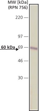
Evaluated by Western Blotting in T-47D cell lysate.
Western Blotting Analysis: A 1:1,000 dilution of this antibody detected Vitamin D3 Receptor in T-47D cell lysate.
Tested applications
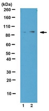
Western Blotting Analysis: A 1:1,000 dilution from a representative lot detected Vitamin D3 Receptor in lysates from Rat liver tissue and HeLa cells.
Affinity Binding Assay: A representative lot of this antibody bound Vitamin D3 Receptor peptide with a KD of 9.5 x 10⁻⁷ in an affinity binding assay.
Immunocytochemistry Analysis: A 1:100 dilution from a representative lot detected Vitamin D3 Receptor in HeLa cells.
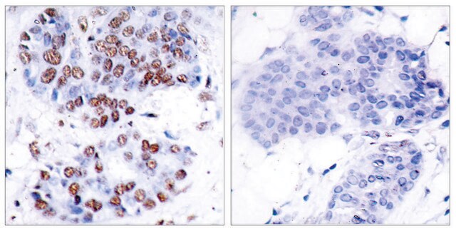
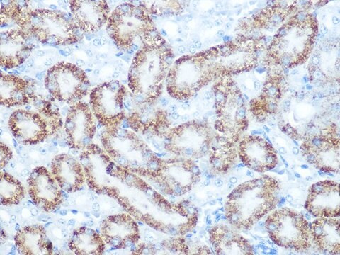
Immunohistochemistry (Paraffin) Analysis: A 1:100 dilution from a representative lot detected Vitamin D3 Receptor in Human small intestine tissue sections.
Note: Actual optimal working dilutions must be determined by end user as specimens, and experimental conditions may vary with the end user
Evaluated by Western Blotting in T-47D cell lysate.
Western Blotting Analysis: A 1:1,000 dilution of this antibody detected Vitamin D3 Receptor in T-47D cell lysate.
표적 설명
Vitamin D3 receptor (UniProt: P11473; also known as VDR, 1,25-dihydroxyvitamin D3 receptor, Nuclear receptor subfamily 1 group I member 1) is encoded by the VDR (also known as NR1I1) gene (Gene ID: 7421) in human. Vitamin D3 receptor (VDR) is a nuclear, ligand-dependent transcription regulator that in complex with hormonally active vitamin D3 regulates the expression of several genes. It is expressed mainly in kidney and intestine and plays a central role in calcium homeostasis. It is mainly localized in the nucleus, but some of it is also found in the cytoplasm. Its nuclear localization is enhanced by vitamin D3. VDR contains three distinct regions, an N-terminal dual zinc finger DNA binding domain, a C-terminal ligand-binding activity domain, and an extensive and unstructured region that links the two functional domains of this protein together. It has two vitamin D3 binding domains (aa 227-237 and 271-278). In the absence of bound vitamin D3 it is present as a homodimer, and it enters the nucleus upon vitamin D3 binding where it forms heterodimers with the retinoid X receptor/RXR. The VDR-RXR heterodimers bind to specific response elements on DNA and activate the transcription of vitamin D3-responsive target genes. Mutations in VDR gene has been linked to a vitamin-D dependent form of Rickets with hypocalcemia and secondary hypoparathyroidism. This ZooMAbZooMAb® recombinant monoclonal antibody, generated by our propriety technology, offers significantly enhanced specificity, affinity, reproducibility, and stability over conventional monoclonals. (Ref.: Kongsbak, M., et al. (2013). Front. Immunol. 4; 148; Pike, JW., and Meyer, MB. (2010). Endocrinol. Metab. Clin. North Am. 39(2): 255-269).
물리적 형태
Purified recombinant rabbit monoclonal antibody IgG, lyophilized in PBS, 5% Trehalose, normal appearance a coarse or translucent resin. The PBS/trehalose components in the ZooMAb formulation can have the appearance of a semi-solid (bead like gel) after lyophilization. This is a normal phenomenon. Please follow the recommended reconstitution procedure in the data sheet to dissolve the semi-solid, bead-like, gel-appearing material. The resulting antibody solution is completely stable and functional as proven by full functional testing. Contains no biocide or preservatives, such as azide, or any animal by-products. Larger pack sizes provided as multiples of 25 μL.
저장 및 안정성
Recommend storage of lyophilized product at 2-8°C; Before reconstitution, micro-centrifuge vials briefly to spin down material to bottom of the vial; Reconstitute each vial by adding 25 μL of filtered lab grade water or PBS; Reconstituted antibodies can be stored at 2-8°C, or -20°C for long term storage. Avoid repeated freeze-thaws.
법적 정보
ZooMAb is a registered trademark of Merck KGaA, Darmstadt, Germany
면책조항
Unless otherwise stated in our catalog or other company documentation accompanying the product(s), our products are intended for research use only and are not to be used for any other purpose, which includes but is not limited to, unauthorized commercial uses, in vitro diagnostic uses, ex vivo or in vivo therapeutic uses or any type of consumption or application to humans or animals.

