추천 제품
생물학적 소스
mouse
항체 형태
purified from hybridoma cell culture
항체 생산 유형
primary antibodies
클론
COL-10, monoclonal
양식
buffered aqueous solution
분자량
~60 kDa
종 반응성
deer, porcine, human
포장
antibody small pack of 25 μL
농도
~1 mg/mL
기술
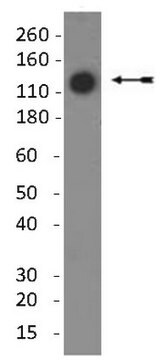
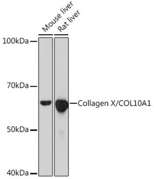
immunoblotting: suitable
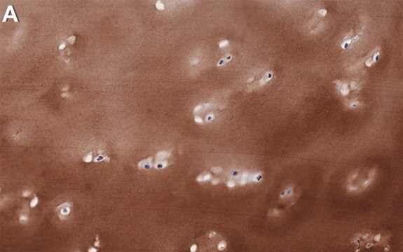
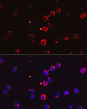
immunofluorescence: 5-10 μg/mL using human osteosarcoma SaOS-2 cells
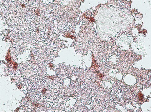
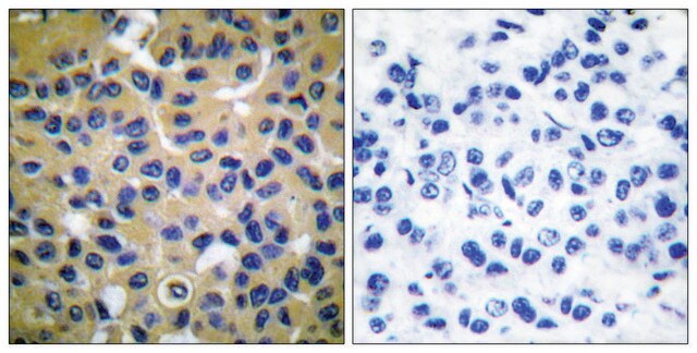
immunohistochemistry: suitable
동형
IgM
UniProt 수납 번호
배송 상태
dry ice
저장 온도
−20°C
타겟 번역 후 변형
unmodified
유전자 정보
human ... COL10A1(1300)
관련 카테고리
일반 설명
The extracellular matrix (ECM) found in the extracellular environment of all tissues and organs, provides the physical microenvironment for cells and a substrate for cell anchorage. It serves as a tissue scaffold and is a dynamic structure whose organization and composition modulate various cellular processes including cell proliferation, attachment, migration, differentiation and survival. The composition of the extracellular framework of all vertebrates is dominated by a Collagen protein family, each member with unique features suited for its function and location.
Type X collagen,also known as Collagen alpha-1(X) chain (COL10A1), is a product of hypertrophic chondrocytes. It shares a similar domain structure with type VIII collagen. In addition, both collagen types represent major components of hexagonal lattice structure, in which the collagen molecules link together by interactions involving the non-triple-helical end regions. Despite these similarities, a distinct tissue distribution has been found for these two molecules: type VIII collagen is distributed in various tissues, whereas type X is restricted to normal fetal hypertrophic cartilage in the growth zones of long bones, vertebrae and ribs and in adult (> 21 yr) thyroid cartilage. It is also found in bone fracture callus, osteoarthritic cartilage and chondrogenic neoplasms, and may be involved in cartilage mineralization. Type X collagen is non-fibrillar, but forms fine pericellular filaments in association with cartilage collagen. It interacts with matrix proteins, such as connexin V, chondrocalcein, collagen II and proteoglycans, as well as with Ca2+ . Mutations in this gene are associated with schmid metaphyseal chondroplasia (MCDS).
The development of antibodies against collagens has provided a powerful method for examining the distribution of these connective tissue proteins and for investigation of epithelial-mesenchymal interactions, tumorigenesis and basement membrane biology in ontogeny and epithelial differentiation.8 Antibodies that react specifically with collagen type X are useful for the study of specific differential tissue expression and the localization of collagen type X.
면역원
Porcine collagen type X
애플리케이션
Monoclonal Anti-Collagen, Type X specifically recognizes native and denatured collagen type X. It does not recognize collagen types I, II, III, V, IX and XI. Reactivity has been observed with human, deer and porcine collagen type X. The antibody is recommended to use in various immunological techniques, including Immunoblotting (∼ 60 kDa in denatured-reduced preparation), Immunofluorescence and Immunohistochemistry.
물리적 형태
Supplied as a solution in 0.01 M phosphate buffered saline pH 7.4, containing 15 mM sodium azide as a preservative.
기타 정보
This product is for R&D use only, not for drug, household, or other uses.
적합한 제품을 찾을 수 없으신가요?
당사의 제품 선택기 도구.을(를) 시도해 보세요.
Storage Class Code
10 - Combustible liquids
WGK
WGK 3
Flash Point (°F)
Not applicable
Flash Point (°C)
Not applicable
가장 최신 버전 중 하나를 선택하세요:
T Aigner et al.
Histochemistry and cell biology, 107(6), 435-440 (1997-06-01)
Little is known about matrix biochemistry and cell differentiation patterns in chondrogenic neoplasms. This is the first description of the focal expression of collagen type X by neoplastic chondrocytes in situ and its incorporation into the extracellular matrix of cartilaginous
G J Rucklidge et al.
Matrix biology : journal of the International Society for Matrix Biology, 15(2), 73-80 (1996-07-01)
Intact collagen type X cannot readily be extracted from the growth plate. Both the use of pepsin to release this molecule from tissue and the relative solubility of collagen type X following treatment of chick embryos with beta-aminopropionitrile (Chen et
I Girkontaite et al.
Matrix biology : journal of the International Society for Matrix Biology, 15(4), 231-238 (1996-09-01)
For studies on processing and tissue distribution of type X collagen, monoclonal antibodies were prepared against human recombinant collagen type X (hrCol X) and tested by ELISA, immunoblotting and immunohistology. Forty-two clones were obtained which were grouped into four different
N Yamaguchi et al.
The Journal of biological chemistry, 264(27), 16022-16029 (1989-09-25)
We have isolated two overlapping cDNA clones covering 2425 base pairs encoding a short type VIII collagen chain synthesized by rabbit corneal endothelial cells. The cDNAs encode an open reading frame of 744 amino acid residues containing a triple-helical domain
Hypertrophic cartilage matrix. Type X collagen, supramolecular assembly, and calcification.
T M Schmid et al.
Annals of the New York Academy of Sciences, 580, 64-73 (1990-01-01)
자사의 과학자팀은 생명 과학, 재료 과학, 화학 합성, 크로마토그래피, 분석 및 기타 많은 영역을 포함한 모든 과학 분야에 경험이 있습니다..
고객지원팀으로 연락바랍니다.