추천 제품
생물학적 소스
goat
결합
unconjugated
항체 형태
affinity isolated antibody
항체 생산 유형
secondary antibodies
클론
polyclonal
형태
buffered aqueous solution
종 반응성
mouse
농도
1.0 mg/mL
기술
immunohistochemistry: suitable
indirect ELISA: suitable
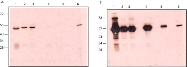
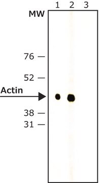
western blot: suitable
배송 상태
wet ice
저장 온도
2-8°C
타겟 번역 후 변형
unmodified
관련 카테고리
일반 설명
Immunoglobulin G (IgG) belongs to the immunoglobulin family and is a widely expressed serum antibody. It consists of a γ heavy chain in the constant (C) region. The monomeric 150kDa structure of IgG constitutes two identical heavy chains and two identical light chains with molecular weight of 50kDa and 25kDa, respectively. The primary structure of this antibody also contains disulfide bonds involved in linking the two heavy chains, linking the heavy and light chains and resides inside the chains. IgG is further subdivided into four classes namely, IgG1, IgG2, IgG3, and IgG4 with different heavy chains, named γ1, γ2, γ3, and γ4, respectively. Limited digestion using papain cleaves the antibody into three fragments, two of which are identical and contain the antigen-binding activity. The third fragment does not possess antigen-binding activity and is known as fragment crystallizable (Fc). It interacts with cells and effector molecules. The Fc fragment contains the CH2 and CH3 domains of the antibody molecule. Pepsin cleaves the carboxy-terminal side of the disulfide bonds in the general region of the antibody and this gives rise to the F(ab′)2 fragment. The two antigen-binding arms of the antibody are linked in this fragment. Maternal IgG is the only antibody transported across the placenta to the fetus. It passively immunizes the infants.
특이성
This product was prepared from monospecific antiserum by immunoaffinity chromatography using Mouse IgG coupled to agarose beads followed by solid phase adsorption(s) to remove any unwanted reactivities, pepsin digestion and chromatographic separation. Assay by immunoelectrophoresis resulted in a single precipitin arc against Anti-Goat Serum, Mouse IgG, Mouse IgG F(c) and Mouse Serum. No reaction was observed against Anti-Pepsin, Anti-Goat IgG F(c), Mouse IgG F(ab′)2 or Bovine, Horse and Human Serum Proteins.
면역원
Mouse IgG F(c) fragment
물리적 특성
Antibody format: IgG F(ab′)2
물리적 형태
Supplied in 0.02 M Potassium Phosphate, 0.15 M Sodium Chloride, pH 7.2
면책조항
Unless otherwise stated in our catalog or other company documentation accompanying the product(s), our products are intended for research use only and are not to be used for any other purpose, which includes but is not limited to, unauthorized commercial uses, in vitro diagnostic uses, ex vivo or in vivo therapeutic uses or any type of consumption or application to humans or animals.
Not finding the right product?
Try our 제품 선택기 도구.
Storage Class Code
10 - Combustible liquids
Flash Point (°F)
Not applicable
Flash Point (°C)
Not applicable
시험 성적서(COA)
제품의 로트/배치 번호를 입력하여 시험 성적서(COA)을 검색하십시오. 로트 및 배치 번호는 제품 라벨에 있는 ‘로트’ 또는 ‘배치’라는 용어 뒤에서 찾을 수 있습니다.
Human placental Fc receptors and the transmission of antibodies from mother to fetus.
Simister NE
Journal of Reproductive Immunology (1997)
Janeway CA
Immunobiology (2001)
Antibody structure, instability, and formulation.
Wang W
Journal of Pharmaceutical Sciences (2007)
Jian Zhang et al.
International journal of oncology, 45(2), 683-690 (2014-06-04)
Tanshinone IIA (TSIIA), a natural diterpene quinone in the traditional Chinese medicinal herb Dan-Shen (Salvia miltiorrhiza), has extensively exerted antitumor activity in cellular and animal models. However, the molecular mechanisms underlying the antitumor effects of TSIIA remain largely unknown. The
Debolina Ghosh et al.
Journal of Alzheimer's disease : JAD, 42(1), 313-324 (2014-05-23)
The extracellular redox environment of cells is mainly set by the redox couple cysteine/cystine (cys/cySS) while intracellular redox is buffered by reduced/oxidized glutathione (GSH/GSSG), but controlled by NAD(P)H/NAD(P). With aging, the extracellular redox environment shifts in the oxidized direction beyond
자사의 과학자팀은 생명 과학, 재료 과학, 화학 합성, 크로마토그래피, 분석 및 기타 많은 영역을 포함한 모든 과학 분야에 경험이 있습니다..
고객지원팀으로 연락바랍니다.


