
HPA042257
Anti-RP1 antibody produced in rabbit
Prestige Antibodies® Powered by Atlas Antibodies, affinity isolated antibody, buffered aqueous glycerol solution
동의어(들):
MAPRE2, Microtubule-associated protein RP/EB family member 2, Anti-Dcdc4a, Anti-Retinitis pigmentosa 1 (autosomal dominant)
로그인조직 및 계약 가격 보기
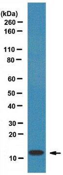
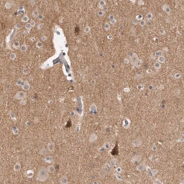
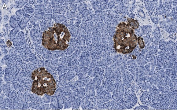
모든 사진(3)
About This Item
추천 제품


생물학적 소스
rabbit
Quality Level
결합
unconjugated
항체 형태
affinity isolated antibody
항체 생산 유형
primary antibodies
클론
polyclonal
제품 라인
Prestige Antibodies® Powered by Atlas Antibodies
양식
buffered aqueous glycerol solution
종 반응성
human
기술
immunohistochemistry: 1:500-1:1000
면역원 서열
VTCSPCEMCTVNKAYSPKETCNPSDTFFPSDGYGVDQTSMNKACFLGEVCSLTDTVFSDKACAQKENHTYEGACPIDETYVPVNVCNTIDFLNSKENTYTDNLDSTEELERGDDIQKDLNILTDPEYKNGFNTLVSHQNVSNLSSCG
UniProt 수납 번호
배송 상태
wet ice
저장 온도
−20°C
타겟 번역 후 변형
unmodified
유전자 정보
human ... RP1(6101)
일반 설명
Retinitis pigmentosa-1 (RP1) is a photoreceptor microtubule-associated protein. RP1 belongs to end binding protein 1(EB1) protein family, which interacts with adenomatous polyposis coli (APC). RP1 is located on human chromosome 8.
면역원
retinitis pigmentosa 1 (autosomal dominant) recombinant protein epitope signature tag (PrEST)
애플리케이션
All Prestige Antibodies Powered by Atlas Antibodies are developed and validated by the Human Protein Atlas (HPA) project and as a result, are supported by the most extensive characterization in the industry.
The Human Protein Atlas project can be subdivided into three efforts: Human Tissue Atlas, Cancer Atlas, and Human Cell Atlas. The antibodies that have been generated in support of the Tissue and Cancer Atlas projects have been tested by immunohistochemistry against hundreds of normal and disease tissues and through the recent efforts of the Human Cell Atlas project, many have been characterized by immunofluorescence to map the human proteome not only at the tissue level but now at the subcellular level. These images and the collection of this vast data set can be viewed on the Human Protein Atlas (HPA) site by clicking on the Image Gallery link. We also provide Prestige Antibodies® protocols and other useful information.
The Human Protein Atlas project can be subdivided into three efforts: Human Tissue Atlas, Cancer Atlas, and Human Cell Atlas. The antibodies that have been generated in support of the Tissue and Cancer Atlas projects have been tested by immunohistochemistry against hundreds of normal and disease tissues and through the recent efforts of the Human Cell Atlas project, many have been characterized by immunofluorescence to map the human proteome not only at the tissue level but now at the subcellular level. These images and the collection of this vast data set can be viewed on the Human Protein Atlas (HPA) site by clicking on the Image Gallery link. We also provide Prestige Antibodies® protocols and other useful information.
생화학적/생리학적 작용
Retinitis pigmentosa-1 (RP1) regulates microtubule formation. Casein kinase II (CK2) a protein kinase when phosphorylates RP1 at Ser(236) aids cell adhesion.RP1 contributes to maintain the architecture of photoreceptor outer segments. Dysfunctioning of RP1 leads to neurodegenerative diseases. Mutations in RP1 gene is known to cause retinitis pigmentosa. Certain regions of RP1 may also cause autosomal recessive rod-cone dystrophy.
특징 및 장점
Prestige Antibodies® are highly characterized and extensively validated antibodies with the added benefit of all available characterization data for each target being accessible via the Human Protein Atlas portal linked just below the product name at the top of this page. The uniqueness and low cross-reactivity of the Prestige Antibodies® to other proteins are due to a thorough selection of antigen regions, affinity purification, and stringent selection. Prestige antigen controls are available for every corresponding Prestige Antibody and can be found in the linkage section.
Every Prestige Antibody is tested in the following ways:
Every Prestige Antibody is tested in the following ways:
- IHC tissue array of 44 normal human tissues and 20 of the most common cancer type tissues.
- Protein array of 364 human recombinant protein fragments.
결합
Corresponding Antigen APREST71374
물리적 형태
Solution in phosphate buffered saline, pH 7.2, containing 40% glycerol and 0.02% sodium azide.
법적 정보
Prestige Antibodies is a registered trademark of Merck KGaA, Darmstadt, Germany
적합한 제품을 찾을 수 없으신가요?
당사의 제품 선택기 도구.을(를) 시도해 보세요.
Storage Class Code
10 - Combustible liquids
WGK
WGK 1
Flash Point (°F)
Not applicable
Flash Point (°C)
Not applicable
가장 최신 버전 중 하나를 선택하세요:
Targeted next generation sequencing identifies novel mutations in RP1 as a relatively common cause of autosomal recessive rod-cone dystrophy
El Shamie, et al.
BioMed Research International, 2015 (2015)
The retinitis pigmentosa 1 protein is a photoreceptor microtubule-associated protein
Liu, Qin, et al.
The Journal of Neuroscience, 24(29), 6427-6436 (2004)
Linkage mapping of autosomal dominant retinitis pigmentosa (RP1) to the pericentric region of human chromosome 8
Blanton,, et al.
Genomics, 11(4), 857-869 (1991)
RP1 is a phosphorylation target of CK2 and is involved in cell adhesion
Stenner,, et al.
PLoS ONE, 8(7), e67595-e67595 (2013)
Molecular basis for photoreceptor outer segment architecture
Goldberg,, et al.
Progress in Retinal and Eye Research, 55, 52-81 (2016)
자사의 과학자팀은 생명 과학, 재료 과학, 화학 합성, 크로마토그래피, 분석 및 기타 많은 영역을 포함한 모든 과학 분야에 경험이 있습니다..
고객지원팀으로 연락바랍니다.