HPA019616
Anti-LPAR2 antibody produced in rabbit
Prestige Antibodies® Powered by Atlas Antibodies, affinity isolated antibody, buffered aqueous glycerol solution
동의어(들):
Anti-LPA receptor 2, Anti-LPA-2, Anti-Lysophosphatidic acid receptor 2, Anti-Lysophosphatidic acid receptor Edg-4
로그인조직 및 계약 가격 보기
모든 사진(1)
About This Item
추천 제품
생물학적 소스
rabbit
결합
unconjugated
항체 형태
affinity isolated antibody
항체 생산 유형
primary antibodies
클론
polyclonal
제품 라인
Prestige Antibodies® Powered by Atlas Antibodies
형태
buffered aqueous glycerol solution
종 반응성
human
기술
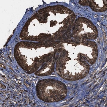
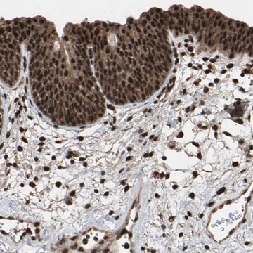
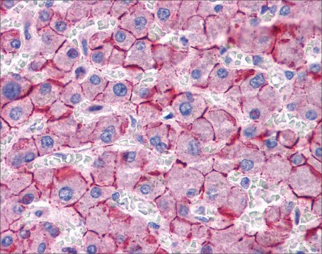
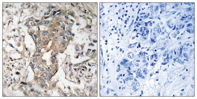
immunohistochemistry: 1:20- 1:50
면역원 서열
LLLDGLGCESCNVLAVEKYFLLLAEANSLVNAAVYSCRDAEMRRTFRRLLCCACLRQSTRESVHYTSSAQGGASTRIMLPENGHPLMDSTL
UniProt 수납 번호
배송 상태
wet ice
저장 온도
−20°C
타겟 번역 후 변형
unmodified
유전자 정보
human ... LPAR2(9170)
일반 설명
LPAR2 (Lysophosphatidic acid receptor 2) is a bioactive lysophospholipid belonging to the endothelial cell differentiation gene (EDG) family of GPCRs. It is widely expressed in different tissues and cell types.
면역원
Lysophosphatidic acid receptor 2 recombinant protein epitope signature tag (PrEST)
애플리케이션
Applications in which this antibody has been used successfully, and the associated peer-reviewed papers, are given below.
Immunohistochemistry (1 paper)
Immunohistochemistry (1 paper)
생화학적/생리학적 작용
LPAR2 (Lysophosphatidic acid receptor 2) is involved in various downstream signaling pathways such as RhoA-ROCK and STAT-3 signaling. It plays a key role in the colorectal cancer (CRC) pathology. In CRC progression it controls cell cycle progression, migration, invasion, and proliferation. During cell migration, it has been reported that cell-cell binding ability depends on the internalization of N-cadherin downstream of lysophosphatidic acid (LPA) receptor 2. LPAR2 is also associated with the receptor-mediated phospholipase C-β3 activation. During activation, C-terminal PDZ domain-binding motif of LPAR2 directly binds to the second PDZ domain of Na(+)/H(+) exchanger regulatory factor2 (NHERF2). Later, that LPAR2 linked PDZ domain of NHERF2 binds to PLC-β3 and forms a complex, which is responsible for gene silencing of PLC-β3.
특징 및 장점
Prestige Antibodies® are highly characterized and extensively validated antibodies with the added benefit of all available characterization data for each target being accessible via the Human Protein Atlas portal linked just below the product name at the top of this page. The uniqueness and low cross-reactivity of the Prestige Antibodies® to other proteins are due to a thorough selection of antigen regions, affinity purification, and stringent selection. Prestige antigen controls are available for every corresponding Prestige Antibody and can be found in the linkage section.
Every Prestige Antibody is tested in the following ways:
Every Prestige Antibody is tested in the following ways:
- IHC tissue array of 44 normal human tissues and 20 of the most common cancer type tissues.
- Protein array of 364 human recombinant protein fragments.
결합
Corresponding Antigen APREST72734
물리적 형태
Solution in phosphate-buffered saline, pH 7.2, containing 40% glycerol and 0.02% sodium azide
법적 정보
Prestige Antibodies is a registered trademark of Merck KGaA, Darmstadt, Germany
면책조항
Unless otherwise stated in our catalog or other company documentation accompanying the product(s), our products are intended for research use only and are not to be used for any other purpose, which includes but is not limited to, unauthorized commercial uses, in vitro diagnostic uses, ex vivo or in vivo therapeutic uses or any type of consumption or application to humans or animals.
적합한 제품을 찾을 수 없으신가요?
당사의 제품 선택기 도구.을(를) 시도해 보세요.
Storage Class Code
10 - Combustible liquids
WGK
WGK 1
Flash Point (°F)
Not applicable
Flash Point (°C)
Not applicable
시험 성적서(COA)
제품의 로트/배치 번호를 입력하여 시험 성적서(COA)을 검색하십시오. 로트 및 배치 번호는 제품 라벨에 있는 ‘로트’ 또는 ‘배치’라는 용어 뒤에서 찾을 수 있습니다.
Ying Zhang et al.
OncoTargets and therapy, 13, 4145-4155 (2020-06-12)
The dysregulation of the human papillomavirus 18 E6 and E7 oncogenes plays a critical role in the angiogenesis of cervical cancer (CC), including the proliferation, migration, and tube formation of vascular endothelial cells. Interfering E6/E7 increases the number of CC
Yong-Seok Oh et al.
Molecular and cellular biology, 24(11), 5069-5079 (2004-05-15)
Lysophosphatidic acid (LPA) activates a family of cognate G protein-coupled receptors and is involved in various pathophysiological processes. However, it is not clearly understood how these LPA receptors are specifically coupled to their downstream signaling molecules. This study found that
Bicheng Yang et al.
BMC women's health, 17(1), 118-118 (2017-11-28)
Given the important roles of the receptor-mediated lysophosphatidic acid (LPA) signaling in both reproductive tract function and gynecological cancers, it will be informative to investigate the potential role of LPA in the development of adenomyosis. The objective of this study
E Sinderewicz et al.
Reproduction in domestic animals = Zuchthygiene, 52(1), 28-34 (2016-09-21)
Lysophosphatidic acid (LPA) exerts various actions on the mammalian reproductive system. In cows, LPA stimulates the synthesis and secretion of luteotropic factors in the ovary, which affects the growth and development of ovarian follicles. The role of LPA in granulosa
Shigeru Hashimoto et al.
Nature communications, 7, 10656-10656 (2016-02-09)
Acquisition of mesenchymal properties by cancer cells is critical for their malignant behaviour, but regulators of the mesenchymal molecular machinery and how it is activated remain elusive. Here we show that clear cell renal cell carcinomas (ccRCCs) frequently utilize the
자사의 과학자팀은 생명 과학, 재료 과학, 화학 합성, 크로마토그래피, 분석 및 기타 많은 영역을 포함한 모든 과학 분야에 경험이 있습니다..
고객지원팀으로 연락바랍니다.