추천 제품
생물학적 소스
rabbit
결합
unconjugated
항체 형태
affinity isolated antibody
항체 생산 유형
primary antibodies
클론
polyclonal
제품 라인
Prestige Antibodies® Powered by Atlas Antibodies
형태
buffered aqueous glycerol solution
종 반응성
human
향상된 검증
independent
RNAi knockdown
Learn more about Antibody Enhanced Validation
기술
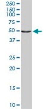
immunoblotting: 0.04-0.4 μg/mL
immunofluorescence: 0.25-2 μg/mL
immunohistochemistry: 1:200-1:500
UniProt 수납 번호
배송 상태
wet ice
저장 온도
−20°C
타겟 번역 후 변형
unmodified
유전자 정보
human ... ACAT1(38)
유사한 제품을 찾으십니까? 방문 제품 비교 안내
면역원
Acetyl-CoA acetyltransferase, mitochondrial precursor recombinant protein epitope signature tag (PrEST)
Sequence
NEQDAYAINSYTRSKAAWEAGKFGNEVIPVTVTVKGQPDVVVKEDEEYKRVDFSKVPKLKTVFQKENGTVTAANASTLNDGAAALVLMTADAAKRLNVTPLARIVAFADAAVEPIDFPIAPVYAASMV
Sequence
NEQDAYAINSYTRSKAAWEAGKFGNEVIPVTVTVKGQPDVVVKEDEEYKRVDFSKVPKLKTVFQKENGTVTAANASTLNDGAAALVLMTADAAKRLNVTPLARIVAFADAAVEPIDFPIAPVYAASMV
애플리케이션
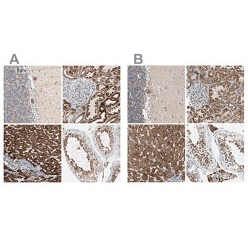
All Prestige Antibodies Powered by Atlas Antibodies are developed and validated by the Human Protein Atlas (HPA) project and as a result, are supported by the most extensive characterization in the industry.
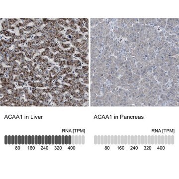
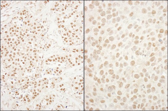
The Human Protein Atlas project can be subdivided into three efforts: Human Tissue Atlas, Cancer Atlas, and Human Cell Atlas. The antibodies that have been generated in support of the Tissue and Cancer Atlas projects have been tested by immunohistochemistry against hundreds of normal and disease tissues and through the recent efforts of the Human Cell Atlas project, many have been characterized by immunofluorescence to map the human proteome not only at the tissue level but now at the subcellular level. These images and the collection of this vast data set can be viewed on the Human Protein Atlas (HPA) site by clicking on the Image Gallery link. We also provide Prestige Antibodies® protocols and other useful information.
The Human Protein Atlas project can be subdivided into three efforts: Human Tissue Atlas, Cancer Atlas, and Human Cell Atlas. The antibodies that have been generated in support of the Tissue and Cancer Atlas projects have been tested by immunohistochemistry against hundreds of normal and disease tissues and through the recent efforts of the Human Cell Atlas project, many have been characterized by immunofluorescence to map the human proteome not only at the tissue level but now at the subcellular level. These images and the collection of this vast data set can be viewed on the Human Protein Atlas (HPA) site by clicking on the Image Gallery link. We also provide Prestige Antibodies® protocols and other useful information.
생화학적/생리학적 작용
ACAT1 (acetyl-CoA acetyltransferase 1) gene encodes a mitochondrial enzyme that is also called as acetoacetyl-CoA thiolase and catalyzes the reversible formation of acetoacetyl-CoA from two molecules of acetyl-CoA. ACAT1 is a key enzyme in the re-utilization of ketone. This protein expression is found to be upregulated in tumor cells for ketone body production and/or utilization and may serve as a potential biomarker for cancer prognosis and a new target for anticancer therapy.
특징 및 장점
Prestige Antibodies® are highly characterized and extensively validated antibodies with the added benefit of all available characterization data for each target being accessible via the Human Protein Atlas portal linked just below the product name at the top of this page. The uniqueness and low cross-reactivity of the Prestige Antibodies® to other proteins are due to a thorough selection of antigen regions, affinity purification, and stringent selection. Prestige antigen controls are available for every corresponding Prestige Antibody and can be found in the linkage section.
Every Prestige Antibody is tested in the following ways:
Every Prestige Antibody is tested in the following ways:
- IHC tissue array of 44 normal human tissues and 20 of the most common cancer type tissues.
- Protein array of 364 human recombinant protein fragments.
결합
Corresponding Antigen APREST86795
물리적 형태
Solution in phosphate-buffered saline, pH 7.2, containing 40% glycerol and 0.02% sodium azide
법적 정보
Prestige Antibodies is a registered trademark of Merck KGaA, Darmstadt, Germany
면책조항
Unless otherwise stated in our catalog or other company documentation accompanying the product(s), our products are intended for research use only and are not to be used for any other purpose, which includes but is not limited to, unauthorized commercial uses, in vitro diagnostic uses, ex vivo or in vivo therapeutic uses or any type of consumption or application to humans or animals.
적합한 제품을 찾을 수 없으신가요?
당사의 제품 선택기 도구.을(를) 시도해 보세요.
Storage Class Code
10 - Combustible liquids
WGK
WGK 1
Flash Point (°F)
Not applicable
Flash Point (°C)
Not applicable
개인 보호 장비
Eyeshields, Gloves, multi-purpose combination respirator cartridge (US)
시험 성적서(COA)
제품의 로트/배치 번호를 입력하여 시험 성적서(COA)을 검색하십시오. 로트 및 배치 번호는 제품 라벨에 있는 ‘로트’ 또는 ‘배치’라는 용어 뒤에서 찾을 수 있습니다.
Howard T Chang et al.
Nutrition & metabolism, 10(1), 47-47 (2013-07-09)
Recent studies in animal models, based on the hypothesis that malignant glioma cells are more dependent on glycolysis for energy generation, have shown promising results using ketogenic diet (KD) therapy as an alternative treatment strategy for malignant glioma, effectively starving
Ubaldo E Martinez-Outschoorn et al.
Cell cycle (Georgetown, Tex.), 11(21), 3964-3971 (2012-10-23)
We have previously proposed that catabolic fibroblasts generate mitochondrial fuels (such as ketone bodies) to promote the anabolic growth of human cancer cells and their metastasic dissemination. We have termed this new paradigm "two-compartment tumor metabolism." Here, we further tested
Sandra Andersson et al.
The journal of histochemistry and cytochemistry : official journal of the Histochemistry Society, 61(11), 773-784 (2013-08-08)
Antibody-based protein profiling on a global scale using immunohistochemistry constitutes an emerging strategy for mapping of the human proteome, which is crucial for an increased understanding of biological processes in the cell. Immunohistochemistry is often performed indirectly using secondary antibodies
Ubaldo E Martinez-Outschoorn et al.
Cell cycle (Georgetown, Tex.), 11(21), 3956-3963 (2012-10-23)
We have previously suggested that ketone body metabolism is critical for tumor progression and metastasis. Here, using a co-culture system employing human breast cancer cells (MCF7) and hTERT-immortalized fibroblasts, we provide new evidence to directly support this hypothesis. More specifically
Charlotte Stadler et al.
Journal of proteomics, 75(7), 2236-2251 (2012-03-01)
We have developed a platform for validation of antibody binding and protein subcellular localization data obtained from immunofluorescence using siRNA technology combined with automated confocal microscopy and image analysis. By combining the siRNA technology with automated sample preparation, automated imaging
자사의 과학자팀은 생명 과학, 재료 과학, 화학 합성, 크로마토그래피, 분석 및 기타 많은 영역을 포함한 모든 과학 분야에 경험이 있습니다..
고객지원팀으로 연락바랍니다.
