
추천 제품


생물학적 소스
goat
결합
FITC conjugate
항체 형태
affinity isolated antibody
항체 생산 유형
secondary antibodies
클론
polyclonal
양식
buffered aqueous solution
기술
direct immunofluorescence: 1:32
저장 온도
2-8°C
타겟 번역 후 변형
unmodified
일반 설명
Human IgGs are glycoprotein antibodies that contain two equivalent light chains and a pair of identical heavy chains. IgGs have four distinct isoforms, ranging from IgG1 to IgG4. These antibodies regulate immunological responses to allergy and pathogenic infections. IgGs have also been implicated in complement fixation and autoimmune disorders
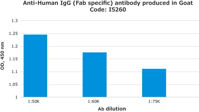
Anti-Human IgG (γ-chain specific), (F(ab′)2) fragment-FITC antibody is specific for human IgG when tested against purified human IgA, IgG, IgM, Bence Jones κ and λ myeloma proteins. The use of this product prevents background staining due to the presence of Fc receptors.
Anti-Human IgG (γ-chain specific), (F(ab′)2) fragment-FITC antibody is specific for human IgG when tested against purified human IgA, IgG, IgM, Bence Jones κ and λ myeloma proteins. The use of this product prevents background staining due to the presence of Fc receptors.
Immunoglobulin G (IgG) belongs to the immunoglobulin family and is a widely expressed serum antibody. The two heavy chains and two light chains of IgG are connected by a disulfide bond. It is a glycoprotein and mainly helps in immune defense. IgG is usually found as a monomer. IgG antibody subtype is the most abundant of serum immunoglobulins of the immune system. It is secreted by B cells and is found in blood and extracellular fluids. About 70 percent of the total immunoglobulin consists of IgG. Immunoglobulin G (IgG) participates in hypersensitivity type II and type III.
면역원
Purified human IgG
애플리케이션
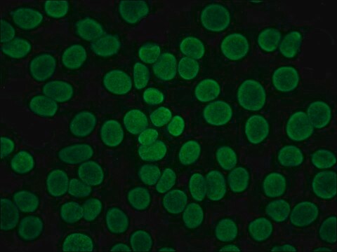
Anti-Human IgG (γ-chain specific), (F(ab′)2) fragment-FITC antibody is suitable for use in direct immunofluorescence (1:32).
Anti-Human IgG (γ-chain specific), F(ab′)2 fragment−FITC antibody has been used in immunofluorescence studies and flow cytometric crossmatch (FCXM).
물리적 형태
Solution in 0.01 M phosphate buffered saline pH 7.4, containing 1% bovine serum albumin and 15 mM sodium azide
면책조항
Unless otherwise stated in our catalog or other company documentation accompanying the product(s), our products are intended for research use only and are not to be used for any other purpose, which includes but is not limited to, unauthorized commercial uses, in vitro diagnostic uses, ex vivo or in vivo therapeutic uses or any type of consumption or application to humans or animals.
적합한 제품을 찾을 수 없으신가요?
당사의 제품 선택기 도구.을(를) 시도해 보세요.
Storage Class Code
12 - Non Combustible Liquids
WGK
nwg
Flash Point (°F)
Not applicable
Flash Point (°C)
Not applicable
이미 열람한 고객


M A Ilham et al.
Transplantation proceedings, 40(6), 1839-1843 (2008-08-05)
Pretransplantation crossmatching is an integral part of kidney transplantation. Flow cytometric crossmatch (FCXM) is more sensitive than complement-dependent cytotoxic crossmatch (CDC-XM). However, the clinical significance of positive FCXM with negative CDC-XM is controversial. We evaluated FCXM in 455 consecutive deceased
Işın Sinem Bağcı et al.
Experimental dermatology, 30(5), 684-690 (2020-12-22)
Ex vivo confocal laser scanning microscopy (CLSM) offers real-time examination of excised tissue in reflectance, fluorescence and digital haematoxylin-eosin (H&E)-like staining modes enabling application of fluorescent-labelled antibodies. We aimed to assess the diagnostic performance of ex vivo CLSM in identifying
Işın S Bağcı et al.
Journal of biophotonics, 12(9), e201800425-e201800425 (2019-04-26)
Ex vivo confocal laser scanning microscopy (ex vivo CLSM) offers an innovative diagnostic approach through vertical scanning of skin samples with a resolution close to conventional histology. In addition, it enables fluorescence detection in tissues. We aimed to assess the
I S Bağcı et al.
Journal of the European Academy of Dermatology and Venereology : JEADV, 33(11), 2123-2130 (2019-07-03)
Ex vivo confocal laser scanning microscopy (ex vivo CLSM) is a novel diagnostic method allowing rapid, high-resolution imaging of excised skin samples. Furthermore, fluorescent detection is possible using fluorescent-labelled antibodies. To assess the applicability of ex vivo CLSM in the
Işın Sinem Bağcı et al.
Acta dermato-venereologica, 97(5), 622-626 (2017-01-18)
Linear IgG deposits along the basement membrane of adnexa has been proposed to be useful in the diagnosis of bullous pemphigoid (BP), but no controlled studies have been performed. This study evaluated linear IgG fluorescence of the basement membrane of
자사의 과학자팀은 생명 과학, 재료 과학, 화학 합성, 크로마토그래피, 분석 및 기타 많은 영역을 포함한 모든 과학 분야에 경험이 있습니다..
고객지원팀으로 연락바랍니다.