
추천 제품

![Monoclonal Anti-CD34-PE antibody produced in mouse clone 4H11[APG], purified immunoglobulin, buffered aqueous solution](/deepweb/assets/sigmaaldrich/product/images/254/307/2ebead45-e77a-4c4f-93bf-0da2c091fe55/640/2ebead45-e77a-4c4f-93bf-0da2c091fe55.jpg)

생물학적 소스
rat
Quality Level
항체 형태
purified antibody
항체 생산 유형
primary antibodies
클론
IM7, monoclonal
제품 라인
ColorWheel®
양식
lyophilized
분자량
calculated mol wt 85.62 kDa
종 반응성
mouse, human
포장
antibody small pack of 25 μL
환경친화적 대안 제품 특성
Waste Prevention
Designing Safer Chemicals
Design for Energy Efficiency
Learn more about the Principles of Green Chemistry.
sustainability
Greener Alternative Product
기술
flow cytometry: suitable
동형
IgG2bκ
에피토프 서열
Unknown
단백질 ID 수납 번호
UniProt 수납 번호
호환성
for use with ColorWheel® Dyes (Required, sold separately)
환경친화적 대안 카테고리
배송 상태
ambient
저장 온도
2-8°C
타겟 번역 후 변형
unmodified
유전자 정보
human ... CD44(12505)
일반 설명
ColorWheel® technology is a novel and proprietary method of creating your own antibody and dye combinations for use in flow cytometry. By incubating any ColorWheel® antibody with any ColorWheel® dye, researchers can quickly and simply produce primary conjugated antibodies for use in single color or multicolor flow cytometry analysis. Each ColorWheel® antibody and dye is lyophilized for long long-term storage, allowing for simplicity and flexibility without compromise. ColorWheel® technology requires both ColorWheel® antibody and ColorWheel® dye for Flow Cytometry application.
We are committed to bringing you greener alternative products, which adhere to one or more of The 12 Principles of Green Chemistry.This product is Preservative-free, lyophilized product for enhanced stability and allow for ambient shipping and thus aligns with "Waste Prevention", "Designing Safer Chemicals" and "Design for Energy Efficiency". Click here for more information.
특이성
Clone IM7 is a rat monoclonal antibody that detects CD44.
면역원
Dexamethasone-induced murine myeloid leukemia M1 cells.
애플리케이션
Quality Control Testing:
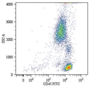
Evaluated by Flow Cytometry in mouse splenoctytes.
Flow Cytometry Analysis (FC): Staining of one million mouse splenocytes was performed using 5 μL of 1:1 mixture of Cat. No. CWA-1002, Anti-Human CD44 (IM7) ColorWheel® Dye-Ready mAb and and Cat. No. CWD-PE ColorWheel® Antibody-Ready Phycoerythrin (PE) Dye or an equivalent amount of PE-conjugated Rat IgG2b isotype control.
Note: Actual optimal working dilutions must be determined by end user as specimens, and experimental conditions may vary with the end user
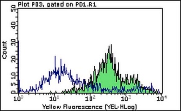
호환성
Requires ColorWheel® Dye (Sold Separately)
표적 설명
CD44 antigen (UniProt: P15379; also known as CDw44, Epican, Extracellular matrix receptor III, ECMR-III, GP90 lymphocyte homing/adhesion receptor, HUTCH-I, Heparan sulfate proteoglycan, Hermes antigen, Hyaluronate receptor, Phagocytic glycoprotein 1, PGP-1, Phagocytic glycoprotein I, PGP-I, CD44) is encoded by the CD44 (also known as LHR, MDU2, MDU3, MIC4) gene (Gene ID: 12505) in murine species. CD44 is a single-pass type I membrane glycoprotein that is synthesized with a signal peptide (aa 1-22), which is subsequently cleaved off to generate the mature form that contains an extracellular domain (aa 23-685), a transmembrane domain (aa 686-707), and a cytoplasmic domain (aa 707-778). It is expressed on all leukocytes, endothelial cells, hepatocytes, and mesenchymal cells. The main ligand for CD44 is Hyaluronic acid (HA) that is expressed by stromal and cancer cells. HA binds the CD44 ligand binding domain and induces conformational changes that allow binding of adaptor proteins or cytoskeletal elements to intracellular domains. This leads to the activation of various signaling pathways leading to cell proliferation, adhesion, migration, and invasion. Overexpression of CD44 has been reported in several cell types and it is considered as an ideal marker for cancer stem cells. (Ref.: Chen, C., et al. (2018). J. Hematol. Oncol. 11; Article 64).
물리적 형태
Lyophilized from PBS containing D-Mannitol and Sucrose, normal appearance is a dried pellet. Reconstituted antibody solution is stable and functional as assessed by functional testing. Contains no biocide or preservatives, such as azide.
재구성
1.0 mg/mL after reconstitution at 25 μL in PBS. Please refer to guidance on suggested starting dilutions and/or titers per application and sample type.
저장 및 안정성
Recommend storage of lyophilized product at 2-8°C. Before reconstitution, micro-centrifuge vials briefly to spin down material to bottom of the vial. Reconstitute each vial by adding 25 μL of PBS. Protect from light.
법적 정보
ColorWheel is a registered trademark of Merck KGaA, Darmstadt, Germany
면책조항
Unless otherwise stated in our catalog or other company documentation accompanying the product(s), our products are intended for research use only and are not to be used for any other purpose, which includes but is not limited to, unauthorized commercial uses, in vitro diagnostic uses, ex vivo or in vivo therapeutic uses or any type of consumption or application to humans or animals.
적합한 제품을 찾을 수 없으신가요?
당사의 제품 선택기 도구.을(를) 시도해 보세요.
관련 제품
제품 번호
설명
가격
Storage Class Code
13 - Non Combustible Solids
WGK
WGK 3
Flash Point (°F)
Not applicable
Flash Point (°C)
Not applicable
가장 최신 버전 중 하나를 선택하세요:
자사의 과학자팀은 생명 과학, 재료 과학, 화학 합성, 크로마토그래피, 분석 및 기타 많은 영역을 포함한 모든 과학 분야에 경험이 있습니다..
고객지원팀으로 연락바랍니다.