추천 제품
제품명
HPCT-05-wt, 12020703
생물학적 소스
human kidney
성장 모드
Adherent
핵형
Not specified
형태학
Epithelial
수용체
Not specified
유사한 제품을 찾으십니까? 방문 제품 비교 안내
세포주 기원
Human proximal tubule cell, kidney, electrolyte transport-competent, SV40 large T-antigen
세포주 설명
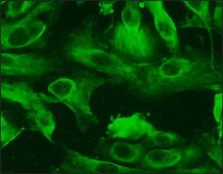
HPCT-05-wt was developed from a primary cultures of proximal tubule epithelial cells from a 50-year old male which were infected with a replication-defective retroviral construct encoding wild-type simian virus 40 large T-antigen. Cells forming electrically resistive monolayers were selected and expanded in culture. HPCT-05-wt cells form polarized, resistive epithelial monolayers with apical microvilli, tight junctional complexes, numerous mitochondria, well-developed Golgi complexes, extensive endoplasmic reticulum, convolutions of the basolateral plasma membrane, and a primary cilium. The HPCT-05-wt cell line exhibits succinate, phosphate, and Na,K-adenosine triphosphatase (ATPase) transport activity, as well as acidic dipeptide- and adenosine triphosphate-regulated mechanisms of ion transport. Transcripts for Na+-bicarbonate cotransporter, Na+-H+ exchanger isoform 3, Na,K-ATPase, parathyroid hormone receptor, epidermal growth factor receptor, and vasopressin V2 receptor were identified. Furthermore, i mMunoreactive sodium phosphate cotransporter type II, vasopressin receptor V1a, and CLIC-1 (NCC27) were also identified. The Y chromosome could not be detected in this cell line by short tandem repeat (STR)-PCR analysis when tested at ECACC. It is a known phenomenon that the Y chromosome can be rearranged or lost in cultured cells resulting in lack of detection. The cell line is identical to the source provided by the depositor based on the STR-PCR analysis. HPCT-05-wt will differentiate at confluence when cultured on a microporous support. The cells should be seeded at 105 cells/cm2 on collagen-coated 30 mM Millicell-CM culture plate inserts (0.4 μm, Millipore) at 37 °C. EGF and serum should be omitted from the apical side of the chamber. On differentiation the cells form polarised resistive epithelial monolayers with apical microvilli, tight junctional complexes, numerous mitochondria, well-developed Golgi complexes, extensive endoplasmic reticulum, convolutions of the basolateral plasma membrane and primary cilium.
DNA 프로파일
STR-PCR Data:
Amelogenin: X
CSF1PO: 10
D13S317: 9
D16S539: 11,13
D5S818: 12,13
D7S820: 9,12
THO1: 6,9.3
TPOX: 8,10
vWA: 14,17
Amelogenin: X
CSF1PO: 10
D13S317: 9
D16S539: 11,13
D5S818: 12,13
D7S820: 9,12
THO1: 6,9.3
TPOX: 8,10
vWA: 14,17
배양 배지
Stemline® Keratinocyte Medium II (S0196) + Stemline® Keratinocyte Growth Supplement (S9945) + L-Glutamine (G7513) + 5 ng/ml human recombinant epidermal growth factor + 5% FBS / FCS (F2442). An alternative medium formulation can be found in Orosz et al., 2004 PMID: 14748622.
계대배양 정규 작업
Split subconfluent cultures (70-80%) 1:3 to 1:6 using 0.25% trypsin/EDTA; 5% CO2; 37 °C. Suggested seeding density 2-4 x 10,000 cells/cm2. Saturation density is approximately 12 x 10,000 cells/cm2. Flasks must be pre-coated with Collagen Type I (C3867) diluted 1:100 with medium without serum. Coating is applied overnight at 37 °C then the flask rinsed with PBS prior to use.
기타 정보
Additional freight & handling charges may be applicable for Asia-Pacific shipments. Please check with your local Customer Service representative for more information.
법적 정보
Stemline is a registered trademark of Merck KGaA, Darmstadt, Germany
가장 최신 버전 중 하나를 선택하세요:
자사의 과학자팀은 생명 과학, 재료 과학, 화학 합성, 크로마토그래피, 분석 및 기타 많은 영역을 포함한 모든 과학 분야에 경험이 있습니다..
고객지원팀으로 연락바랍니다.