
추천 제품


생물학적 소스
mouse
결합
unconjugated
항체 형태
purified immunoglobulin
항체 생산 유형
primary antibodies
클론
CL0296, monoclonal
제품 라인
Prestige Antibodies® Powered by Atlas Antibodies
양식
buffered aqueous glycerol solution
종 반응성
human
향상된 검증
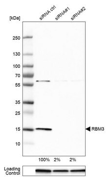
orthogonal RNAseq
RNAi knockdown
Learn more about Antibody Enhanced Validation
기술
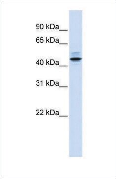
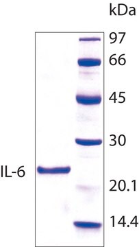
immunoblotting: 1 μg/mL
immunofluorescence: 2-10 μg/mL
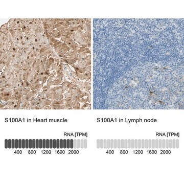
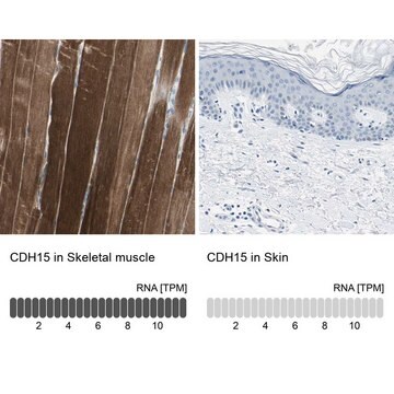
immunohistochemistry: 1:500- 1:1000
동형
IgG1
Ensembl | 인체 수납 번호
UniProt 수납 번호
배송 상태
wet ice
저장 온도
−20°C
타겟 번역 후 변형
unmodified
유전자 정보
human ... RBM3(5935)
면역원
Recombinant protein corresponding to RNA binding motif (RNP1, RRM) protein 3.
Sequence
DEQALEDHFSSFGPISEVVVVKDRETQRSRGFGFITFTNPEHASVAMRAMNGESLDGRQIRVDHAGKSARGTRGGGFGAHGRGRSYSRGGGDQGYGSGRYYDSRPGGYGYGYGRSRDYNGRNQGGYDRYSGGNY
Sequence
DEQALEDHFSSFGPISEVVVVKDRETQRSRGFGFITFTNPEHASVAMRAMNGESLDGRQIRVDHAGKSARGTRGGGFGAHGRGRSYSRGGGDQGYGSGRYYDSRPGGYGYGYGRSRDYNGRNQGGYDRYSGGNY
애플리케이션
All Prestige Antibodies Powered by Atlas Antibodies are developed and validated by the Human Protein Atlas (HPA) project and as a result, are supported by the most extensive characterization in the industry.
The Human Protein Atlas project can be subdivided into three efforts: Human Tissue Atlas, Cancer Atlas, and Human Cell Atlas. The antibodies that have been generated in support of the Tissue and Cancer Atlas projects have been tested by immunohistochemistry against hundreds of normal and disease tissues and through the recent efforts of the Human Cell Atlas project, many have been characterized by immunofluorescence to map the human proteome not only at the tissue level but now at the subcellular level. These images and the collection of this vast data set can be viewed on the Human Protein Atlas (HPA) site by clicking on the Image Gallery link. We also provide Prestige Antibodies® protocols and other useful information.
The Human Protein Atlas project can be subdivided into three efforts: Human Tissue Atlas, Cancer Atlas, and Human Cell Atlas. The antibodies that have been generated in support of the Tissue and Cancer Atlas projects have been tested by immunohistochemistry against hundreds of normal and disease tissues and through the recent efforts of the Human Cell Atlas project, many have been characterized by immunofluorescence to map the human proteome not only at the tissue level but now at the subcellular level. These images and the collection of this vast data set can be viewed on the Human Protein Atlas (HPA) site by clicking on the Image Gallery link. We also provide Prestige Antibodies® protocols and other useful information.
특징 및 장점
Prestige Antibodies® are highly characterized and extensively validated antibodies with the added benefit of all available characterization data for each target being accessible via the Human Protein Atlas portal linked just below the product name at the top of this page. The uniqueness and low cross-reactivity of the Prestige Antibodies® to other proteins are due to a thorough selection of antigen regions, affinity purification, and stringent selection. Prestige antigen controls are available for every corresponding Prestige Antibody and can be found in the linkage section.
Every Prestige Antibody is tested in the following ways:
Every Prestige Antibody is tested in the following ways:
- IHC tissue array of 44 normal human tissues and 20 of the most common cancer type tissues.
- Protein array of 364 human recombinant protein fragments.
결합
Corresponding Antigen APREST74363
물리적 형태
Phospate buffered saline, pH 7.2, containing 40% glycerol and 0.02% sodium azide
법적 정보
Prestige Antibodies is a registered trademark of Merck KGaA, Darmstadt, Germany
면책조항
Unless otherwise stated in our catalog or other company documentation accompanying the product(s), our products are intended for research use only and are not to be used for any other purpose, which includes but is not limited to, unauthorized commercial uses, in vitro diagnostic uses, ex vivo or in vivo therapeutic uses or any type of consumption or application to humans or animals.
적합한 제품을 찾을 수 없으신가요?
당사의 제품 선택기 도구.을(를) 시도해 보세요.
Storage Class Code
10 - Combustible liquids
WGK
WGK 1
Flash Point (°F)
Not applicable
Flash Point (°C)
Not applicable
가장 최신 버전 중 하나를 선택하세요:
RBM3-regulated genes promote DNA integrity and affect clinical outcome in epithelial ovarian cancer.
Åsa Ehlén et al.
Translational oncology, 4(4), 212-221 (2011-08-02)
The RNA-binding motif protein 3 (RBM3) was initially discovered as a putative cancer biomarker based on its differential expression in various cancer forms in the Human Protein Atlas (HPA). We previously reported an association between high expression of RBM3 and
Björn Nodin et al.
Diagnostic pathology, 7, 82-82 (2012-07-19)
Malignant melanoma is the most lethal form of skin cancer with a variable clinical course even in patients with thin melanomas and localized disease. Despite increasing insights into melanoma biology, no prognostic biomarkers have yet been incorporated into clinical protocols.
Barbara Hjelm et al.
Proteomics. Clinical applications, 5(11-12), 624-635 (2011-10-01)
In this study, we investigated the prognostic impact of human RBM3 expression in colorectal cancer using tissue microarray-based immunohistochemical analysis. One polyclonal antibody and four monoclonal anti-RBM3 antibodies were generated and epitope mapped using two different methods. Bacterial display revealed
Spatiotemporal profile and essential role of RBM3 expression after spinal cord injury in adult rats.
Zhiming Cui et al.
Journal of molecular neuroscience : MN, 54(2), 252-263 (2014-03-29)
Hypoxia and other adverse conditions are usually encountered by rapidly growing cells. The RNA-binding motif protein 3 (RBM3) is induced by low temperature and hypoxia. However, its expression and function in spinal cord injury are still unclear. To investigate the
자사의 과학자팀은 생명 과학, 재료 과학, 화학 합성, 크로마토그래피, 분석 및 기타 많은 영역을 포함한 모든 과학 분야에 경험이 있습니다..
고객지원팀으로 연락바랍니다.