추천 제품
생물학적 소스
rabbit
Quality Level
100
500
결합
unconjugated
항체 형태
culture supernatant
항체 생산 유형
primary antibodies
클론
EP184, monoclonal
설명
For In Vitro Diagnostic Use in Select Regions (See Chart)
양식
buffered aqueous solution
종 반응성
human
포장
vial of 0.1 mL concentrate (408R-14)
vial of 0.5 mL concentrate (408R-15)
bottle of 1.0 mL predilute (408R-17)
vial of 1.0 mL concentrate (408R-16)
bottle of 7.0 mL predilute (408R-18)
제조업체/상표
Cell Marque™
기술
immunohistochemistry (formalin-fixed, paraffin-embedded sections): 1:25-1:100
동형
IgG
제어
kidney, renal oncocytoma
배송 상태
wet ice
저장 온도
2-8°C
시각화
cytoplasmic, nuclear
유전자 정보
human ... S100A1(6271)
일반 설명
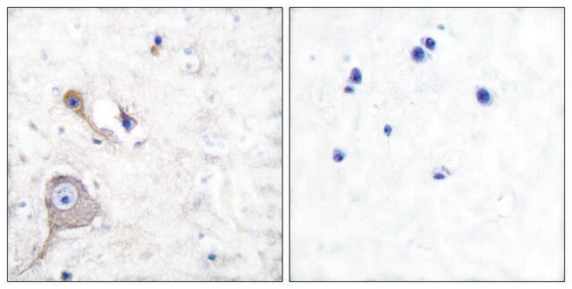
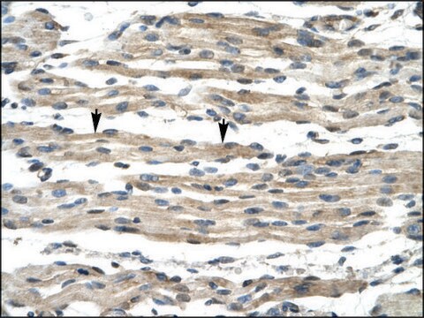
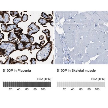
S100A1 is a member of S100 protein family of calcium binding proteins.S100A1 is very useful in the identification of renal cell carcinoma.Published literature indicates that 37 of 40 renal oncocytomas (93%), including two with the tubulo-cystic growth pattern, reacted positively by anti-S100A1.Most of them showed both cytoplasmic and nuclear immunostaining with moderate-to-strong intensity. S100A1 immunostaining was also observed in 30 of 41 clear cell renal cell carcinomas (73%); 6 showed focal positivity, whereas 24 displayed an immunoreactivity ranging from moderate (12 cases) to diffuse (12 cases) expression. Anti-S100A1 detected 30 cases of 32 papillary renal cell carcinomas (94%), 6 showing focal, 12 moderate, and 12 diffuse immunoreactivity. This study also demonstrated that 48 of the 51 chromophobe renal cell carcinomas (94%) showed no labeling by anti-S100A1. In 3 cases of chromophobe renal cell carcinomas (6%) that were positive by anti-S100A1, 2 were classic variant showing scattered S100A1 immunoreactive cells and an eosinophilic variant with nucleocytoplasmic staining in about 40% of the neoplastic cells.Therefore, anti-S100A1 is very useful in the differentiation of renal oncocytoma from chromophobe renal cell carcinoma as well as distinguishing clear cell renal cell carcinoma from chromophobe renal cell carcinoma. S100A1 is detected in normal renal tissue and expressed in both the nuclei and the cytoplasm of the cells lining proximal tubules, loops of Henle, and collecting ducts. The glomerular components are negative. S100A1 is also expressed by skeletal muscle and myocardium; and specific cytoplasmic expression is found in follicular dendritic cells of lymph node.
품질
 IVD |  IVD |  IVD |  RUO |
결합
S100A1 Positive Control Slides, Product No. 408S, are available for immunohistochemistry (formalin-fixed, paraffin-embedded sections).
물리적 형태
Solution in Tris Buffer, pH 7.3-7.7, with 1% BSA and <0.1% Sodium Azide
제조 메모
Download the IFU specific to your product lot and formatNote: This requires a keycode which can be found on your packaging or product label.
기타 정보
For Technical Service please contact: 800-665-7284 or email: service@cellmarque.com
법적 정보
Cell Marque is a trademark of Merck KGaA, Darmstadt, Germany
적합한 제품을 찾을 수 없으신가요?
당사의 제품 선택기 도구.을(를) 시도해 보세요.
Storage Class Code
12 - Non Combustible Liquids
WGK
WGK 2
Flash Point (°F)
Not applicable
Flash Point (°C)
Not applicable
가장 최신 버전 중 하나를 선택하세요:
시험 성적서(COA)
Lot/Batch Number
Paolo Cossu Rocca et al.
Modern pathology : an official journal of the United States and Canadian Academy of Pathology, Inc, 20(7), 722-728 (2007-05-08)
S100A1 is a calcium-binding protein, which has been recently found in renal cell neoplasms. We evaluated the diagnostic utility of immunohistochemical detection of S100A1 in 164 renal cell neoplasms. Forty-one clear cell, 32 papillary, and 51 chromophobe renal cell carcinomas
D B Zimmer et al.
Brain research bulletin, 37(4), 417-429 (1995-01-01)
The S100 family of calcium binding proteins contains approximately 16 members each of which exhibits a unique pattern of tissue/cell type specific expression. Although the distribution of these proteins is not restricted to the nervous system, the implication of several
자사의 과학자팀은 생명 과학, 재료 과학, 화학 합성, 크로마토그래피, 분석 및 기타 많은 영역을 포함한 모든 과학 분야에 경험이 있습니다..
고객지원팀으로 연락바랍니다.


