
추천 제품
생물학적 소스
mouse
Quality Level
100
500
결합
unconjugated
항체 형태
culture supernatant
항체 생산 유형
primary antibodies
클론
16/f5, monoclonal
설명
For In Vitro Diagnostic Use in Select Regions (See Chart)
양식
buffered aqueous solution
종 반응성
human
포장
vial of 0.1 mL concentrate (376M-94)
vial of 0.5 mL concentrate (376M-95)
bottle of 1.0 mL predilute (376M-97)
vial of 1.0 mL concentrate (376M-96)
bottle of 7.0 mL predilute (376M-98)
제조업체/상표
Cell Marque™
기술
immunohistochemistry (formalin-fixed, paraffin-embedded sections): 1:100-1:500
동형
IgG1κ
제어
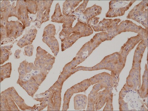
breast, pancreatic ductal adenocarcinoma, urothelial carcinoma
배송 상태
wet ice
저장 온도
2-8°C
시각화
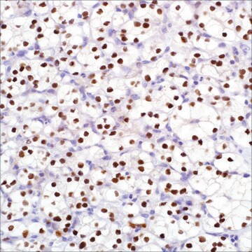
cytoplasmic, nuclear
유전자 정보
human ... S100P(6286)
일반 설명
S100P is a member of the S100 family of proteins. The family is expressed in a wide range of cells and is thought to play a role in cell cycle progression and in differentiation. S100P was initially identified in the placenta at rather high levels. Anti-S100P with nuclear or nuclear/ cytoplasmic immunoreactivity can be seen in essentially 100% of pancreatic ductal adenocarcinoma in pancreatic resection, and fine needle aspiration biopsy specimens. Anti-S100P displays no staining in the benign pancreatic ducts and acinar glands. S100P has been detected in the cells of virtually all intraductal papillary mucinous neoplasms tested. S100P is clearly expressed in the invasive component of intraductal papillary mucinous neoplasms (100%), including perineural, lymphatic, and minimal invasion. Biopsies of bile ducts with primary adenocarcinomas (90%) have exhibited strong nuclear and cytoplasmic staining for anti-S100P, with none of the 32 benign biopsies exhibiting anti-S100P immunoreactivity. An immunohistochemical panel including anti-S100P can be helpful in distinguishing adenocarcinoma from reactive epithelial changes on challenging bile duct biopsies. The detection of S100P expression may help distinguish urothelial carcinomas from other genitourinary neoplasms that enter into the differential diagnosis.
품질
 IVD |  IVD |  IVD |  RUO |
결합
S100P Positive Control Slides, Product No. 376S, are available for immunohistochemistry (formalin-fixed, paraffin-embedded sections).
물리적 형태
Solution in Tris Buffer, pH 7.3-7.7, with 1% BSA and <0.1% Sodium Azide
제조 메모
Download the IFU specific to your product lot and formatNote: This requires a keycode which can be found on your packaging or product label.
기타 정보
For Technical Service please contact: 800-665-7284 or email: service@cellmarque.com
법적 정보
Cell Marque is a trademark of Merck KGaA, Darmstadt, Germany
적합한 제품을 찾을 수 없으신가요?
당사의 제품 선택기 도구.을(를) 시도해 보세요.
가장 최신 버전 중 하나를 선택하세요:
시험 성적서(COA)
Lot/Batch Number
Tatjana Crnogorac-Jurcevic et al.
The Journal of pathology, 201(1), 63-74 (2003-09-02)
In order to expand our understanding of the molecular changes underlying the complex pathology of pancreatic malignancy, global gene expression profiling of pancreatic adenocarcinoma compared with normal pancreatic tissue was performed. Human cDNA arrays comprising 9932 elements were interrogated with
Kohei Nakata et al.
Human pathology, 41(6), 824-831 (2010-02-16)
Intraductal papillary mucinous neoplasms of the pancreas are subclassified based on morphological features, and different immunohistochemical profiles have been identified in association with the subtypes. We previously reported that S100P was an early developmental marker of pancreatic carcinogenesis and that
Mary Levy et al.
Human pathology, 41(9), 1210-1219 (2010-04-13)
Histopathologic distinction between benign and malignant bile duct epithelial lesions on endoscopic biopsies can be extremely challenging because of limited material, crush artifact, and compounding inflammatory and/or reactive changes particularly after stent placement. In this study, a total of 72
John P T Higgins et al.
The American journal of surgical pathology, 31(5), 673-680 (2007-04-27)
The morphologic distinction between prostate and urothelial carcinoma can be difficult. To identify novel diagnostic markers that may aid in the differential diagnosis of prostate versus urothelial carcinoma, we analyzed expression patterns in prostate and bladder cancer tissues using complementary
Hongbing Deng et al.
American journal of clinical pathology, 129(1), 81-88 (2007-12-20)
Even though the cytologic criteria for pancreatic ductal adenocarcinoma (PDA) on fine-needle aspiration biopsy (FNAB) specimens have been well defined, a diagnostic challenge is still present. We immunohistochemically evaluated the diagnostic value of S100P on cell-block and/or smear preparations in
자사의 과학자팀은 생명 과학, 재료 과학, 화학 합성, 크로마토그래피, 분석 및 기타 많은 영역을 포함한 모든 과학 분야에 경험이 있습니다..
고객지원팀으로 연락바랍니다.