콘텐츠로 건너뛰기
Merck
모든 사진(1)

주요 문서

356M-1

Sigma-Aldrich

Cytokeratin 5 & 6 (D5 & 16B4) Mouse Monoclonal Antibody

로그인조직 및 계약 가격 보기


About This Item

UNSPSC 코드:
12352203
NACRES:
NA.41

생물학적 소스

mouse

Quality Level

100
500

결합

unconjugated

항체 형태

culture supernatant

항체 생산 유형

primary antibodies

클론

D5 & 16B4, monoclonal

설명

For In Vitro Diagnostic Use in Select Regions (See Chart)

양식

buffered aqueous solution

종 반응성

human

포장

vial of 0.1 mL concentrate (356M-14)
vial of 0.5 mL concentrate (356M-15)
bottle of 1.0 mL predilute (356M-17)
vial of 1.0 mL concentrate (356M-16)
bottle of 7.0 mL predilute (356M-18)

제조업체/상표

Cell Marque

기술

immunohistochemistry (formalin-fixed, paraffin-embedded sections): 1:50-1:200

동형

IgG1

제어

mesothelioma

배송 상태

wet ice

저장 온도

2-8°C

시각화

cytoplasmic

유전자 정보

human ... KRT5(3852)

일반 설명

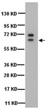
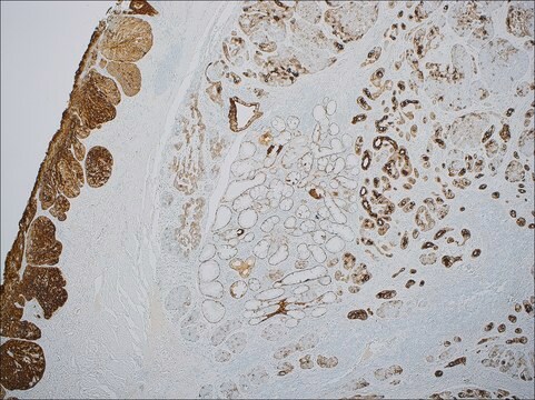
Anti-CK 5 & 6 positivity is seen in 65% to 100% of epithelioid mesotheliomas, in 0% to 19% of lung adenocarcinomas, and in 22% to 80% of serous carcinoma of the ovary and peritoneum. Keratin 5 & 6 expression in lung adenocarcinoma is not common, and when it occurs, the reaction is usually confined to small focal areas or scattered cells. The most probable cause of this finding is the presence of squamous differentiation of lung adenocarcinoma. Anti-CK 5 & 6 positivity has been useful in recognizing squamous cell carcinoma of the skin. Less than 10% of carcinomas of the breast, colon, and prostate stain positively for this marker. Anti-CK 5 & 6 has also been used successfully as a myoepithelial cell marker in the prostate and breast to determine malignancy. Anti-CK 5 & 6 is a useful marker to distinguish lung squamous cell carcinoma from lung adenocarcinoma and large cell carcinoma within a panel including antibodies against TTF-1, Napsin A, p63, SOX-2, desmocollin3, and desmoglein3.

품질


IVD

IVD

IVD

RUO

결합

Cytokeratin 5 & 6 Positive Control Slides, Product No. 356S, are available for immunohistochemistry (formalin-fixed, paraffin-embedded sections).

물리적 형태

Solution in Tris Buffer, pH 7.3-7.7, with 1% BSA and <0.1% Sodium Azide

제조 메모

Download the IFU specific to your product lot and formatNote: This requires a keycode which can be found on your packaging or product label.

기타 정보

For Technical Service please contact: 800-665-7284 or email: service@cellmarque.com

법적 정보

Cell Marque is a trademark of Merck KGaA, Darmstadt, Germany

적합한 제품을 찾을 수 없으신가요?  

당사의 제품 선택기 도구.을(를) 시도해 보세요.


가장 최신 버전 중 하나를 선택하세요:

시험 성적서(COA)

Lot/Batch Number

적합한 버전을 찾을 수 없으신가요?

특정 버전이 필요한 경우 로트 번호나 배치 번호로 특정 인증서를 찾을 수 있습니다.

이 제품을 이미 가지고 계십니까?

문서 라이브러리에서 최근에 구매한 제품에 대한 문서를 찾아보세요.

문서 라이브러리 방문

Magali Lacroix-Triki et al.
Virchows Archiv : an international journal of pathology, 442(6), 548-554 (2003-04-25)
Previous studies have shown that basal-type cytokeratins (CKs) can distinguish usual ductal hyperplasia (UDH) from the spectrum of atypical ductal hyperplasia (ADH), ductal carcinoma in situ (DCIS) and lobular carcinoma in situ (LCIS). Indeed, expression of these CKs is weak
Jorge S Reis-Filho et al.
Virchows Archiv : an international journal of pathology, 443(2), 122-132 (2003-07-29)
p63, cytokeratin (CK) 5/6 and CK 14 have been employed in diagnostic pathology as markers of basal, squamous and myoepithelial differentiation in several types of human neoplasms; however, there is scant data on the concurrent expression of these markers in
Peiguo G Chu et al.
Modern pathology : an official journal of the United States and Canadian Academy of Pathology, Inc, 15(1), 6-10 (2002-01-18)
Cytokeratin 5/6 (CK 5/6) immunoreactivity has been observed in the vast majority of cases of malignant mesothelioma but only rarely in pulmonary adenocarcinomas. Thus, CK 5/6 has been used to distinguish malignant mesothelioma from pulmonary adenocarcinoma. However, the utility of
J E Sigel et al.
Journal of cutaneous pathology, 28(10), 520-524 (2001-12-12)
Cutaneous spindle cell squamous cell carcinoma (SSCC) is a challenging diagnosis since it may be difficult to distinguish from spindle cell melanoma, leiomyosarcoma and atypical fibroxanthoma. Furthermore, it may be difficult to demonstrate epithelial differentiation by a traditional immunohistochemical panel.
Lin Lin et al.
Journal of cutaneous pathology, 30(2), 114-117 (2003-03-19)
Epithelioid sarcoma (ES) is a rare, aggressive soft tissue tumor characterized by nodular aggregates of epithelioid and/or spindled cells that are immunoreactive to cytokeratins (CKs) and epithelial membrane antigen. ES that arises in the dermis may cause epidermal ulceration and

관련 콘텐츠

Diagnostic immunohistochemistry overview highlights its importance in cancer diagnosis and detecting infectious diseases in modern clinical pathology.

자사의 과학자팀은 생명 과학, 재료 과학, 화학 합성, 크로마토그래피, 분석 및 기타 많은 영역을 포함한 모든 과학 분야에 경험이 있습니다..

고객지원팀으로 연락바랍니다.