추천 제품
생물학적 소스
rabbit
Quality Level
100
500
결합
unconjugated
항체 형태
Ig fraction of antiserum
항체 생산 유형
primary antibodies
클론
polyclonal
설명
For In Vitro Diagnostic Use in Select Regions (See Chart)
양식
buffered aqueous solution
종 반응성
human
포장
vial of 0.1 mL concentrate (267A-14)
vial of 0.5 mL concentrate (267A-15)
bottle of 1.0 mL predilute (267A-17)
vial of 1.0 mL concentrate (267A-16)
bottle of 7.0 mL predilute (267A-18)
제조업체/상표
Cell Marque®
기술
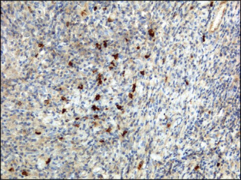
immunohistochemistry (formalin-fixed, paraffin-embedded sections): 1:100-1:500
제어
tonsil
배송 상태
wet ice
저장 온도
2-8°C
시각화
cytoplasmic
일반 설명
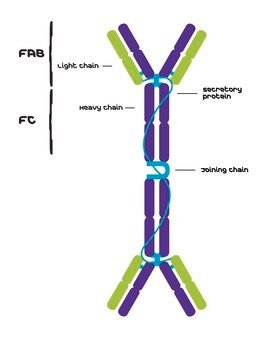
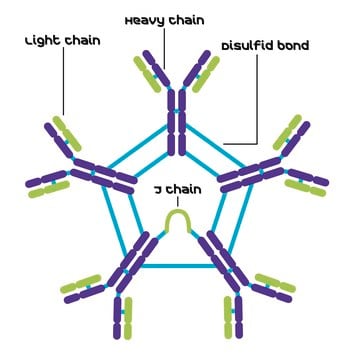
Anti-IgA antibody reacts with surface immunoglobulin IgA alpha chains. It is useful when identifying leukemias, plasmacytomas, and B-cell lineage derived Hodgkin′s lymphomas. Due to the restricted expression of heavy and light chains in these diseases, demonstration of B-cell lymphoma/plasmacytoma is aided with this antibody.
품질
 IVD |  IVD |  IVD |  RUO |
결합
IgA (polyclonal) Positive Control Slides, Product No. 267S, are available for immunohistochemistry (formalin-fixed, paraffin-embedded sections).
물리적 형태
Solution in Tris Buffer, pH 7.3-7.7, with 1% BSA and <0.1% Sodium Azide
제조 메모
Download the IFU specific to your product lot and formatNote: This requires a keycode which can be found on your packaging or product label.
기타 정보
For Technical Service please contact: 800-665-7284 or email: service@cellmarque.com
법적 정보
Cell Marque is a registered trademark of Merck KGaA, Darmstadt, Germany
적합한 제품을 찾을 수 없으신가요?
당사의 제품 선택기 도구.을(를) 시도해 보세요.
가장 최신 버전 중 하나를 선택하세요:
시험 성적서(COA)
Lot/Batch Number
Tissue section immunologic methods in lymphomas
Warnake, R., et al.
Diagnostic Immunohistochemistry (Masson Publishing), 203-221 (1981)
Manual of Diagnostic Antibodies for Immunohistology, 217-219 (1999)
A Arnold et al.
The New England journal of medicine, 309(26), 1593-1599 (1983-12-29)
Immunoglobulin genes in their germ-line form are separated DNA subsegments that must be joined by means of recombinations during B-cell development. Individual immunoglobulin-gene rearrangements are specific for a given B cell and its progeny. We show that the detection of
Diffuse polyclonal B-cell lymphoma during primary infection with Epstein-Barr virus.
J E Robinson et al.
The New England journal of medicine, 302(23), 1293-1297 (1980-06-05)
C R Taylor
Archives of pathology & laboratory medicine, 102(3), 113-121 (1978-03-01)
Immunoperoxidase methods have much in common with established immunofluorescence procedures. Both have the potential for specific demonstration of cell and tissue antigens, with similar limitations demanding rigorous control of specificity. In any study the choice of an immunofluorescence method or
자사의 과학자팀은 생명 과학, 재료 과학, 화학 합성, 크로마토그래피, 분석 및 기타 많은 영역을 포함한 모든 과학 분야에 경험이 있습니다..
고객지원팀으로 연락바랍니다.