추천 제품
생물학적 소스
mouse
Quality Level
100
500
결합
unconjugated
항체 형태
diluted ascites fluid
항체 생산 유형
primary antibodies
클론
C8/144B, monoclonal
설명
For In Vitro Diagnostic Use in Select Regions (See Chart)
형태
buffered aqueous solution
종 반응성
human
포장
vial of 0.1 mL concentrate (108M-94)
vial of 0.5 mL concentrate (108M-95)
bottle of 1.0 mL predilute (108M-97)
vial of 1.0 mL concentrate (108M-96)
bottle of 7.0 mL predilute (108M-98)
제조업체/상표
Cell Marque™
기술
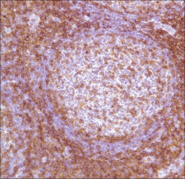
immunohistochemistry (formalin-fixed, paraffin-embedded sections): 1:25-1:100
동형
IgG1κ
제어
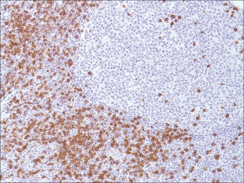
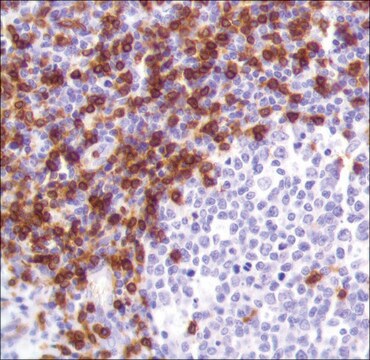
tonsil
배송 상태
wet ice
저장 온도
2-8°C
시각화
membranous
유전자 정보
human ... CD8A(925)
일반 설명
Anti-CD8 is a T cell marker for the detection of cytotoxic/suppresser cells of blood lymphocytes. CD8 is also detected on NK cells, most thymocytes, a subpopulation of null cells and bone marrow cells. This antibody, along with other markers, can be used to distinguish between reactive and neoplastic T-cells.
품질
 IVD |  IVD |  IVD |  RUO |
결합
CD8 Positive Control Slides, Product No. 108S, are available for immunohistochemistry (formalin-fixed, paraffin-embedded sections).
물리적 형태
Solution in Tris Buffer, pH 7.3-7.7, with 1% BSA and <0.1% Sodium Azide
제조 메모
Download the IFU specific to your product lot and formatNote: This requires a keycode which can be found on your packaging or product label.
기타 정보
For Technical Service please contact: 800-665-7284 or email: service@cellmarque.com
법적 정보
Cell Marque is a trademark of Merck KGaA, Darmstadt, Germany
적합한 제품을 찾을 수 없으신가요?
당사의 제품 선택기 도구.을(를) 시도해 보세요.
시험 성적서(COA)
제품의 로트/배치 번호를 입력하여 시험 성적서(COA)을 검색하십시오. 로트 및 배치 번호는 제품 라벨에 있는 ‘로트’ 또는 ‘배치’라는 용어 뒤에서 찾을 수 있습니다.
M L Rossi et al.
Journal of clinical pathology, 41(3), 314-319 (1988-03-01)
Twenty four meningiomas (17 benign and seven "atypical" were reacted with a panel of monoclonal antibodies to macrophages, lymphocytes, and HLA DR antigens. All the tumours contained macrophages but these cells were more numerous in the atypical meningiomas. Lymphocytes, almost
Immunohistological analysis of human lymphoma: correlation of histological and immunological categories.
H Stein et al.
Advances in cancer research, 42, 67-147 (1984-01-01)
F Phan-Dinh-Tuy et al.
Molecular immunology, 19(12), 1649-1654 (1982-12-01)
Three human lymphocyte differentiation antigens, specific of the entire T-cell population, of the helper/inducer T-cell subset, and of the cytotoxic/suppressor T-cell subset have been identified, using mouse monoclonal antibodies obtained from Dr. P. Kung. Various T-cell populations were radio-labelled, the
J D Nuckols et al.
Journal of cutaneous pathology, 26(4), 169-175 (1999-05-21)
Differentiation between mycosis fungoides (MF) and cutaneous inflammatory processes can usually be made on clinical and histologic grounds. In difficult cases, immunohistochemical studies can be helpful since MF infiltrates usually contain a predominance of CD4+ lymphocytes, while most inflammatory lesions
D Y Mason et al.
Journal of clinical pathology, 45(12), 1084-1088 (1992-12-01)
To evaluate whether cytotoxic/suppressor T cells can be detected in paraffin wax embedded human tissue samples using antibodies to a synthetic CD8 peptide sequence. Polyclonal and monoclonal antibodies were raised against a 13 amino acid peptide sequence from the cytoplasmic
자사의 과학자팀은 생명 과학, 재료 과학, 화학 합성, 크로마토그래피, 분석 및 기타 많은 영역을 포함한 모든 과학 분야에 경험이 있습니다..
고객지원팀으로 연락바랍니다.