추천 제품
생물학적 소스
rabbit
Quality Level
항체 형태
culture supernatant
항체 생산 유형
primary antibodies
클론
Vli37, monoclonal
종 반응성
bovine, mouse
종 반응성(상동성에 의해 예측)
rat (based on 100% sequence homology)
기술
immunocytochemistry: suitable
immunohistochemistry: suitable (paraffin)
western blot: suitable
동형
IgG
NCBI 수납 번호
UniProt 수납 번호
배송 상태
dry ice
타겟 번역 후 변형
unmodified
유전자 정보
mouse ... Cdh5(12562)
일반 설명
Cadherin-5 (UniProt P55284; also known as Vascular endothelial cadherin, VE-Cad, VE-cadherin, VEcad, CD144) is encoded by the Cdh5 gene (Gene ID 12562) in murine species. Cadherins (calcium-dependent adhesion) are type-1 transmembrane proteins that form adherens junctions in tissues and play important roles in mediating cell adhesion. Cadherins are designated with a prefix that specifies their tissue association. Vascular endothelial-cadherin (VE-cadherin) is the transmembrane component of the endothelial adherens junction between vascular endothelial cells (ECs) and plays a pivotal role in endothelium integrity and in the control of vascular permeability. One characteristic of VEGF-induced vascular permeability is the phosphorylation of VE-cadherin, which leads to VE-cadherin internalization and the destabilization of adherens junctions. Likewise, VE-cadherin overexpression is shown to decrease the permeability of endothelial monolayers in vitro. VE-cadherin is initially produced with a signal peptide (a.a. 1-24) and a propeptide (a.a. 25-45) sequence, the removal of which yields tthe mature protein containing a large extracellular region (a.a. 46-599) with five cadherine repeats (a.a. 46-149, 150-256, 257-371, 372-476, 477-593), followed by a transmembrane segment (a.a. 600-620) and a cytoplasmic domain (a.a. 621-784).
특이성
Clone Vli37 stains endothelial adherens junctions by immunocytochemistry and immunohistochemistry.
면역원
Epitope: Near C-terminus.
Linear peptide corresponding to the C-terminal cytoplasmic domain sequence of mouse VE-Cadherin.
애플리케이션
Research Category
Cell Structure
Cell Structure
Research Sub Category
Adhesion (CAMs)
Adhesion (CAMs)
This Anti-VE-Cadherin Antibody, clone Vli37 is validated for use in Western Blotting, Immunohistochemistry (Paraffin), Immunocytochemistry for the detection of VE-Cadherin.
Western Blotting Analysis: An 1:500 dilution of this antibody detected VE-Cadherin in 10 µg of mouse brain endothelial bEnd.3 cell lysate.
Western Blotting Analysis: An 1:2,000 dilution from a representative lot detected VE-cadherin expression in lysastes from mouse lung tissue, mouse brain endothelial bEnd.3 cells, and bovine aortic endothelial GM7372 cells (Courtesy of Dr. Volkhard Lindner, Maine Medical Center Research Institute, Scarborough, ME).
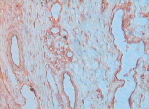
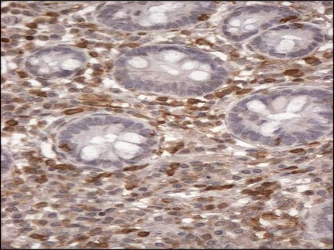
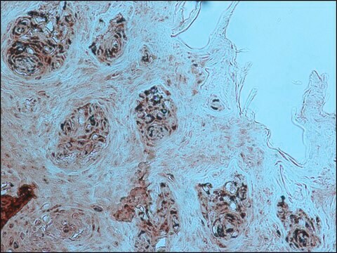
Immunohistochemistry Analysis: An 1:250-5,000 dilution from a representative lot detected VE-cadherin immunoreactivity in formalin-fixed, paraffin-embeded mouse lung, aorta, and liver tissue sections (Courtesy of Dr. Volkhard Lindner, Maine Medical Center Research Institute, Scarborough, ME).
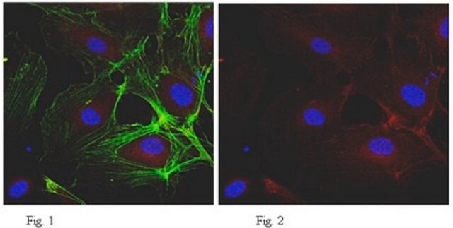
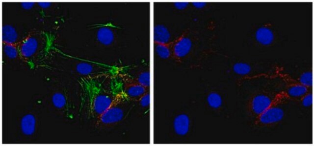
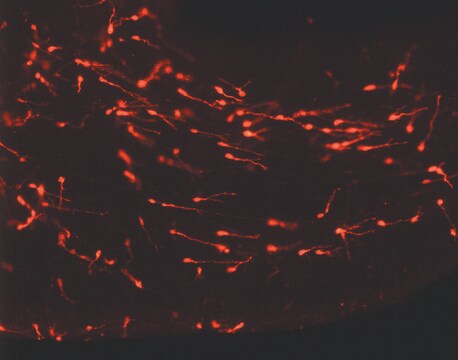
Immunocytochemistry Analysis: An 1:500-5,000 dilution from a representative lot detected VE-cadherin immunoreactivity in bovine aortic endothelial GM7372 cells (Courtesy of Dr. Volkhard Lindner, Maine Medical Center Research Institute, Scarborough, ME).
품질
Evaluated by Western Blotting in mouse lung tissue lysate.
Western Blotting Analysis: An 1:500 dilution of this antibody detected VE-Cadherin in 10 µg of mouse lung tissue lysate.
Western Blotting Analysis: An 1:500 dilution of this antibody detected VE-Cadherin in 10 µg of mouse lung tissue lysate.
표적 설명
~110 observed. Target band size appears larger than the calculated molecular weight of 83.05 kDa due to glycosylation.
물리적 형태
Rabbit monoclonal IgG in supernatant with 0.05% sodium azide.
Unpurified
저장 및 안정성
Stable for 1 year at -20°C from date of receipt.
Handling Recommendations: Upon receipt and prior to removing the cap, centrifuge the vial and gently mix the solution. Aliquot into microcentrifuge tubes and store at -20°C. Avoid repeated freeze/thaw cycles, which may damage IgG and affect product performance.
Handling Recommendations: Upon receipt and prior to removing the cap, centrifuge the vial and gently mix the solution. Aliquot into microcentrifuge tubes and store at -20°C. Avoid repeated freeze/thaw cycles, which may damage IgG and affect product performance.
기타 정보
Concentration: Please refer to lot specific datasheet.
면책조항
Unless otherwise stated in our catalog or other company documentation accompanying the product(s), our products are intended for research use only and are not to be used for any other purpose, which includes but is not limited to, unauthorized commercial uses, in vitro diagnostic uses, ex vivo or in vivo therapeutic uses or any type of consumption or application to humans or animals.
적합한 제품을 찾을 수 없으신가요?
당사의 제품 선택기 도구.을(를) 시도해 보세요.
Storage Class Code
10 - Combustible liquids
WGK
WGK 2
시험 성적서(COA)
제품의 로트/배치 번호를 입력하여 시험 성적서(COA)을 검색하십시오. 로트 및 배치 번호는 제품 라벨에 있는 ‘로트’ 또는 ‘배치’라는 용어 뒤에서 찾을 수 있습니다.
Chi Zhang et al.
Arteriosclerosis, thrombosis, and vascular biology, 38(11), 2691-2705 (2018-10-26)
Objective- Blood-CNS (central nervous system) barrier defects are implicated in retinopathies, neurodegenerative diseases, stroke, and epilepsy, yet, the pathological mechanisms downstream of barrier defects remain incompletely understood. Blood-retina barrier (BRB) formation and retinal angiogenesis require β-catenin signaling induced by the
자사의 과학자팀은 생명 과학, 재료 과학, 화학 합성, 크로마토그래피, 분석 및 기타 많은 영역을 포함한 모든 과학 분야에 경험이 있습니다..
고객지원팀으로 연락바랍니다.