
추천 제품


생물학적 소스
rat
Quality Level
항체 형태
purified antibody
항체 생산 유형
primary antibodies
클론
PMab-1, monoclonal
종 반응성
mouse
포장
antibody small pack of 25 μg
기술
flow cytometry: suitable
immunocytochemistry: suitable
immunohistochemistry: suitable (paraffin)
western blot: suitable
동형
IgG2aκ
NCBI 수납 번호
UniProt 수납 번호
타겟 번역 후 변형
unmodified
유전자 정보
mouse ... Pdpn(14726)
일반 설명
Podoplanin (UniProt: Q62011; also known as Aggrus, Glycoprotein 38, Gp38, OTS-8, PA2.26 antigen, T1-alpha, T1A, Transmembrane glycoprotein E11) I encoded by the Pdpn (also known as Gp38, Ots8) gene (Gene ID: 14726) in murine species. Podoplanin serves as the endogenous ligand of C-type lectin-like receptor-2 (CLEC-2) and is highly expressed in various tumors and in some normal cells, such as lymphatic endothelial cells and podocytes. Podoplanin is detected at high levels in lung and brain, at lower levels in kidney, stomach, liver, spleen and esophagus, and not detected in skin and small intestine. It is expressed in epithelial cells of choroid plexus, ependyma, glomerulus and alveolus, in mesothelial cells and in endothelia of lymphatic vessels. It is also expressed in stromal cells of peripheral lymphoid tissue and thymic epithelial cells and is detected in carcinoma cell lines and cultured fibroblasts. High expression of Podoplanin is observed in colon carcinomas. . It may be involved in cell migration and/or actin cytoskeleton organization. Podoplanin is localized to actin-rich microvilli and plasma membrane projections, such as filopodia, lamellipodia, and ruffles. Extensively O-glycosylated. Contains sialic acid residues. O-glycosylation is necessary for platelet aggregation activity. Podoplanin levels are down-regulated by treatment with puromycin aminonucleoside. Mice lacking or with defective podoplanin die at birth of respiratory failure due to a low number of attenuated type I cells, narrow and irregular air spaces, and defective formation of alveolar saccules. Administration of PMab-1 antibody is shown to reduce lymphangiogenesis in the corneal suture and ear wound healing murine models. It suppresses thioglycollate-induced macrophages infiltration at the site of wound healing. (Ref.: Maruyama, Y et al (2014). Invest.Ophthal. Visual Sci. 55(8); 4813-4822).
특이성
Clone PMab-1 detects murine Podoplanin. It targets an epitipe within 14 amino acids from the N-terminal region.
면역원
KLH-conjugated linear peptide corresponding to 14 amino acids from the N-terminal region of murine Podoplanin.
애플리케이션
Anti-Podoplanin, clone PMab-1, Cat. No. MABT851, is a rat monoclonal antibody that detects mouse Podoplanin and has been tested for use in Flow Cytometry, Immunocytochemistry, Immunohistochemistry (Paraffin), and Western Blotting.
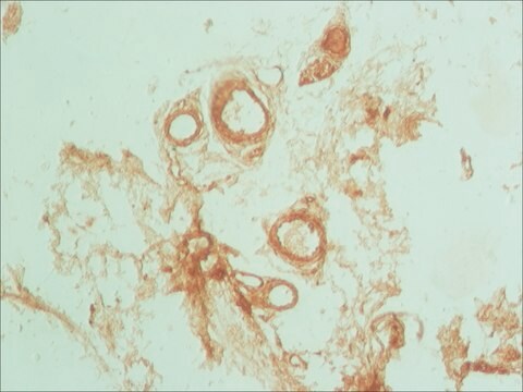
Immunohistochemistry Analysis: A 1:50 dilution from a representative lot detected Podoplanin in mouse lung tissue.
Immunohistochemistry Analysis: A representative lot detected Podoplanin in Immunohistochemistry applications (Kaji, C., et. al. (2012) Acta Histochem Cytochem. 45(4):227-37).
Flow Cytometry Analysis: A representative lot detected Podoplanin in Flow Cytometry applications (Oki, H., et. al. (2015). Monoclon Antib Immunodiagn Immunother. 34(6):396-403).
Western Blotting Analysis: A representative lot detected Podoplanin in Western Blotting applications (Oki, H., et. al. (2015). Monoclon Antib Immunodiagn Immunother. 34(6):396-403).
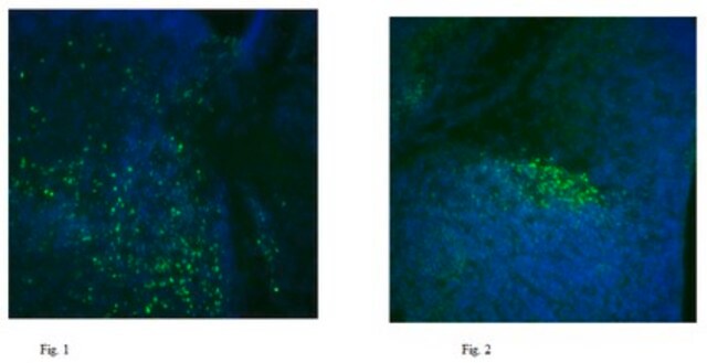
Immunocytochemistry Analysis: A representative lot detected Podoplanin in Immunocytochemistry applications (Kaji, C., et. al. (2012) Acta Histochem Cytochem. 45(4):227-37).
Immunohistochemistry Analysis: A representative lot detected Podoplanin in Immunohistochemistry applications (Kaji, C., et. al. (2012) Acta Histochem Cytochem. 45(4):227-37).
Flow Cytometry Analysis: A representative lot detected Podoplanin in Flow Cytometry applications (Oki, H., et. al. (2015). Monoclon Antib Immunodiagn Immunother. 34(6):396-403).
Western Blotting Analysis: A representative lot detected Podoplanin in Western Blotting applications (Oki, H., et. al. (2015). Monoclon Antib Immunodiagn Immunother. 34(6):396-403).
Immunocytochemistry Analysis: A representative lot detected Podoplanin in Immunocytochemistry applications (Kaji, C., et. al. (2012) Acta Histochem Cytochem. 45(4):227-37).
Research Category
Cell Structure
Cell Structure
품질
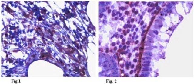
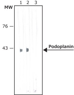
Evaluated by Western Blotting in mouse colon tissue lysate.
Western Blotting Analysis: 1 µg/mL of this antibody detected Podoplanin in 10 µg of mouse colon tissue lysate.
Western Blotting Analysis: 1 µg/mL of this antibody detected Podoplanin in 10 µg of mouse colon tissue lysate.
표적 설명
~42 kDa observed; 18.23 kDa calculated. Uncharacterized bands may be observed in some lysate(s).
물리적 형태
Format: Purified
Protein G purified
Purified rat monoclonal antibody IgG2a in buffer containing 0.1 M Tris-Glycine (pH 7.4), 150 mM NaCl with 0.05% sodium azide.
저장 및 안정성
Stable for 1 year at 2-8°C from date of receipt.
기타 정보
Concentration: Please refer to lot specific datasheet.
면책조항
Unless otherwise stated in our catalog or other company documentation accompanying the product(s), our products are intended for research use only and are not to be used for any other purpose, which includes but is not limited to, unauthorized commercial uses, in vitro diagnostic uses, ex vivo or in vivo therapeutic uses or any type of consumption or application to humans or animals.
적합한 제품을 찾을 수 없으신가요?
당사의 제품 선택기 도구.을(를) 시도해 보세요.
Storage Class Code
12 - Non Combustible Liquids
WGK
WGK 1
Flash Point (°F)
Not applicable
Flash Point (°C)
Not applicable
시험 성적서(COA)
제품의 로트/배치 번호를 입력하여 시험 성적서(COA)을 검색하십시오. 로트 및 배치 번호는 제품 라벨에 있는 ‘로트’ 또는 ‘배치’라는 용어 뒤에서 찾을 수 있습니다.
자사의 과학자팀은 생명 과학, 재료 과학, 화학 합성, 크로마토그래피, 분석 및 기타 많은 영역을 포함한 모든 과학 분야에 경험이 있습니다..
고객지원팀으로 연락바랍니다.