MABT844
Anti-Cytokeratin 8/18 Antibody, clone L2A1
clone L2A1, from mouse
동의어(들):
Cytokeratin-8/18, Keratin type II cytoskeletal 8/Keratin type I cytoskeletal 18, Keratin-8/Keratin-18, K8/K18)
로그인조직 및 계약 가격 보기
모든 사진(1)
About This Item
UNSPSC 코드:
12352203
eCl@ss:
32160702
NACRES:
NA.43
클론:
L2A1, monoclonal
application:
ICC
IP
IP
종 반응성:
human
기술:
immunocytochemistry: suitable
immunoprecipitation (IP): suitable
immunoprecipitation (IP): suitable
citations:
5
추천 제품
생물학적 소스
mouse
항체 형태
purified antibody
항체 생산 유형
primary antibodies
클론
L2A1, monoclonal
종 반응성
human
포장
antibody small pack of 25 μg
기술
immunocytochemistry: suitable
immunoprecipitation (IP): suitable
동형
IgGκ
타겟 번역 후 변형
unmodified
유전자 정보
human ... KRT18(3875)
일반 설명
Cytokeratin-8/18 (UniProt: P05787/P05783; also known as Keratin, type II cytoskeletal 8/ Keratin, type I cytoskeletal 18, Keratin-8/Keratin-18, K8/K18) is encoded by the KRT8/KRT18 (also known as CYK8/CYK18) gene (Gene ID: 3856/3875) in human. Cytokeratins 8 and 18 (K8/18) are simple epithelial cell-specific intermediate filament proteins. Cytokeratin-8 is a type II, neutral to basic, protein of the intermediate filament family that together with Cytokeratin-19 (KRT19) helps to link the contractile apparatus to dystrophin at the costameres of striated muscle. Cytokeratin-8 can undergo phosphorylation on three major serine residues: Serine 23, 431, and 73. Serine 23 is shown to be highly conserved in all type II keratins. Phosphorylation at Serine 73 is reported to increase during cellular stress, including hear and drug exposure. However, under normal conditions serine 73 remains largely dephosphorylated. Cytokeratin-8 can also undergo O-glycosylation in a cell cycle-dependent manner and glycosylation increases its solubility and reduces stability by inducing proteasomal degradation. Cytokeratin-18 is a type I, acidic, protein that is expressed in colon, placenta, and liver. Higher expression levels have been reported in lymph nodes of breast carcinoma. Cytokeratin-18 is involved in the uptake of thrombin-antithrombin complexes by hepatic cells. Upon phosphorylation, it plays a role in filament reorganization. It is proteolytically cleaved by caspases during epithelial cell apoptosis and this cleavage is shown to occur at Asp-238. Phosphorylation of cytokeratin-18 at serine 33 is shown to increase during mitosis in cultured cells and in regenerating liver. Serine 33 phosphorylation is considered to be essential for its association with 14-3-3 proteins and has a role in keratin organization and distribution. Cytokeratin-18 can undergo O-glycosylation, which results in its increased solubility and reduction in its stability. Mutations in KRT8 and KRT18 genes have been been linked to liver cirrhosis that is characterized by severe panlobular liver-cell swelling with Mallory body formation, prominent pericellular fibrosis, and marked deposits of copper.
특이성
Clone L2A1 is a mouse monoclonal antibody that detects human cytokeratin 8/18.
면역원
KLH-conjugated Linear Peptide corresponding to a sequence from Human Cytokeratin 8/18.
애플리케이션
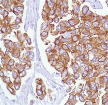
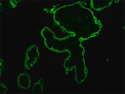
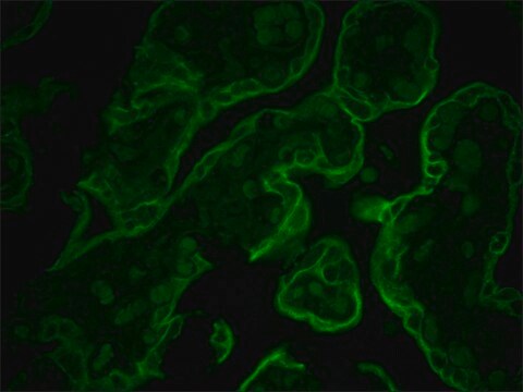
Anti-Cytokeratin 8/18, clone L2A1, Cat. No. MABT844, is a mouse monoclonal antibody that detects cytokeratin 8/18 and has been tested for use in Immunocytochemistry and Immunoprecipitation.
Immunoprecipitation Analysis: A representative lot detected Cytokeratin 8/18 in Immunoprecipitation applications (Haltiwanger, R.S., et. al. (1997). J Biol Chem. 272(13):8752-8; Chou, C.F., et. al. (1994). Biochem J. 298 ( Pt 2):457-63; Chou, C.F., et. al. (1993). Biochem J. 268(6):4465-72; Ku, N.O., et. al. (2000). J Cell Biol. 149(3):547-52; Lahdeniemi, I.A.K., et. al. (2017). Cell Death Differ. 24(6):984-996).
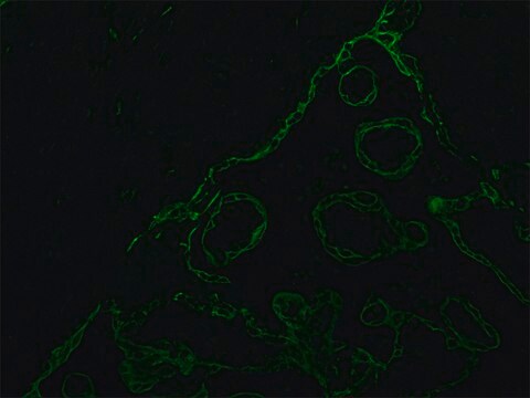
Immunocytochemistry Analysis: A representative lot detected Cytokeratin 8/18 in Immunocytochemistry applications (Chou, C.F., et. al. (1993). Biochem J. 268(6):4465-72).
Immunocytochemistry Analysis: A representative lot detected Cytokeratin 8/18 in Immunocytochemistry applications (Chou, C.F., et. al. (1993). Biochem J. 268(6):4465-72).
Research Category
Cell Structure
Cell Structure
품질
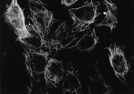
Evaluated by Immunocytochemistry in HT-29 cells.
Immunocytochemistry Analysis: A 1:500 dilution of this antibody detected Cytokeratin 8/18 in HT-29 cells.
Immunocytochemistry Analysis: A 1:500 dilution of this antibody detected Cytokeratin 8/18 in HT-29 cells.
표적 설명
~53.70 kDa and 48.06 kDa, respectively for cytokeratin 8 and 18 calculated. Uncharacterized bands may be observed in some lysate(s).
물리적 형태
Format: Purified
Protein G purified
Purified mouse monoclonal antibody IgG in buffer containing 0.1 M Tris-Glycine (pH 7.4), 150 mM NaCl with 0.05% sodium azide.
저장 및 안정성
Stable for 1 year at 2-8°C from date of receipt.
기타 정보
Concentration: Please refer to lot specific datasheet.
면책조항
Unless otherwise stated in our catalog or other company documentation accompanying the product(s), our products are intended for research use only and are not to be used for any other purpose, which includes but is not limited to, unauthorized commercial uses, in vitro diagnostic uses, ex vivo or in vivo therapeutic uses or any type of consumption or application to humans or animals.
적합한 제품을 찾을 수 없으신가요?
당사의 제품 선택기 도구.을(를) 시도해 보세요.
시험 성적서(COA)
제품의 로트/배치 번호를 입력하여 시험 성적서(COA)을 검색하십시오. 로트 및 배치 번호는 제품 라벨에 있는 ‘로트’ 또는 ‘배치’라는 용어 뒤에서 찾을 수 있습니다.
C F Chou et al.
The Biochemical journal, 298 ( Pt 2), 457-463 (1994-03-01)
We describe the characterization of an acidic glycoprotein (molecular mass approximately 85 kDa) that associates with keratin intermediate filaments of 'simple'-type epithelia. Using a number of anti-keratin monoclonal antibodies, the 85 kDa glycoprotein was identified by co-immunoprecipitation with keratin polypeptides
N O Ku et al.
The Journal of cell biology, 149(3), 547-552 (2000-05-03)
Keratin polypeptides 8 and 18 (K8/18) are intermediate filament (IF) proteins that are expressed in glandular epithelia. Although the mechanism of keratin turnover is poorly understood, caspase-mediated degradation of type I keratins occurs during apoptosis and the proteasome pathway has
C F Chou et al.
The Journal of biological chemistry, 268(6), 4465-4472 (1993-02-25)
Arrest of the human colonic cell line HT29 at the G2/M phase of the cell cycle resulted in changes in keratin assembly that were coupled with a significant increase in the O-linked glycosylation and serine phosphorylation of keratin polypeptides 8
R S Haltiwanger et al.
The Journal of biological chemistry, 272(13), 8752-8758 (1997-03-28)
O-Linked N-acetylglucosamine (O-GlcNAc) is a ubiquitous and abundant protein modification found on nuclear and cytoplasmic proteins. Several lines of evidence suggest that it is a highly dynamic modification and that the levels of this sugar on proteins may be regulated.
Iris A K Lähdeniemi et al.
Cell death and differentiation, 24(6), 984-996 (2017-05-06)
Keratins (K) are intermediate filament proteins important in stress protection and mechanical support of epithelial tissues. K8, K18 and K19 are the main colonic keratins, and K8-knockout (K8-/-) mice display a keratin dose-dependent hyperproliferation of colonic crypts and a colitis-phenotype.
자사의 과학자팀은 생명 과학, 재료 과학, 화학 합성, 크로마토그래피, 분석 및 기타 많은 영역을 포함한 모든 과학 분야에 경험이 있습니다..
고객지원팀으로 연락바랍니다.