추천 제품
생물학적 소스
mouse
Quality Level
항체 형태
purified immunoglobulin
항체 생산 유형
primary antibodies
클론
SC-71, monoclonal
종 반응성
human, opossum, rat, mouse
반응하면 안 됨
guinea pig
종 반응성(상동성에 의해 예측)
bovine (based on 100% sequence homology)
기술
immunofluorescence: suitable
immunohistochemistry: suitable
western blot: suitable
동형
IgG1κ
NCBI 수납 번호
UniProt 수납 번호
배송 상태
ambient
타겟 번역 후 변형
unmodified
유전자 정보
human ... MYH2(4620)
일반 설명
Myosin-2 (UniProt Q9BE41; also known as MyHC-2a, MyHC-IIa, Myosin heavy chain 2, Myosin heavy chain 2a, Myosin heavy chain IIa, Myosin heavy chain, skeletal muscle, adult 2) is encoded by the MYHSA gene (Gene ID 788772) in bovine species. Myosin heavy chain (MyHC) is a major structural component of the striated muscle contractile apparatus and is essential for body movement and cardiac contractility. MyHC are encoded by a highly conserved multigene family, of which eight isoforms have been identified in mammals, each encoded by a separate gene that displays distinct temporal-spatial regulation. MyHC-2a is expressed in the fast-type 2A muscle fibers. Recessive MyHC-2a myopathy caused by missense mutations results in mild muscle weakness. Autosomal dominant mutation (E706K) is reported to cause defective muscle function and compromises the structural integrity of all muscle cells. Ref.: Tajsharghi, H et al. (2014). Eur. J. Hum. Genet. 22: 801-808.
특이성
Clone SC-71 immunostained type 2A, but not type 1, 2B, or 2X, fibers in rat tibialis anterior muscle by targeting an epitope within the light meromyosin region (Schiaffino, S., et al. (1989). J. Muscle Res. Cell Motil. 10(3):197-205).
면역원
Bovine skeletal muscle myosin (Schiaffino, S., et al. (1989). J. Muscle Res. Cell Motil. 10(3):197-205).
애플리케이션
Research Category
Cell Structure
Cell Structure
This mouse monoclonal Anti-Myosin-2 (MYH2) Antibody, clone SC-71, Cat. No. MABT840, is validated for use in Immunofluorescence, Immunohistochemistry, and Western Blotting for the detection of Myosin-2.
Western Blotting Analysis: 0.5 µg/mL from a representative lot detected Myosin-2 (MYH2) in 10 µg of human gastrocnemious muscle and showed minimal reactivity with soleus embryonic muscle tissue lysates (Courtesy of Alberto Rossi, Ph.D., University of Colorado, U.S.A.).
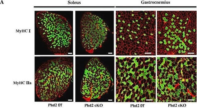
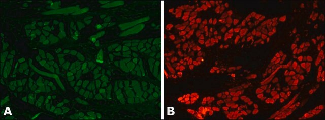
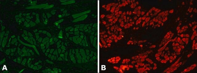
Immunofluorescence Analysis: A representative lot immunostained type 2A fibers in mouse hindlimb muscle cryosections encompassing soleus and surrounding tissues (Kurapati, R., et al. (2012). Hum. Mol. Genet. 21(8):1706-1724).
Immunofluorescence Analysis: A representative lot immunostained type 2A fibers in rat soleus muscle cryosections following Bupivacaine-induced muscle regeneration. In tetrodotoxin/TTX-paralyzed-regenerated muscles type 2A MHC was not expressed (Midrio, M., et al. (2002). Basic Appl. Myol. 12(2): 77-80).
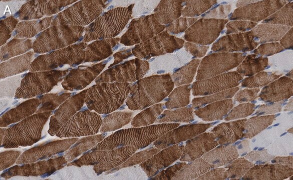
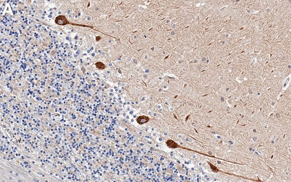
Immunohistochemistry Analysis: A representative lot detected type IIA myosin heavy chain (MyHC) in human masseter (jaw) muscle cryosections (Horton, M.J., et al. (2001). Arch. Oral Biol. 46(11):1039-1050).
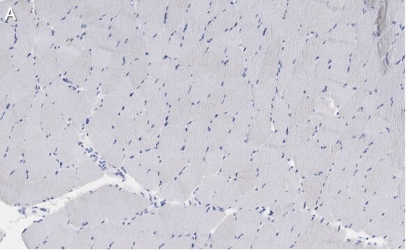
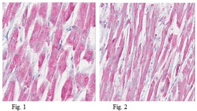
Immunohistochemistry Analysis: Representative lots immunostained type 2A, but not type 1, 2B, or 2X, fibers in soleus (rat) and tibialis (mouse, rat, and Mgray short-tailed opossum/Monodelphis domestica) anterior muscle cryosections. Clone SC-71 failed to stain guinea pig tibialis sections (Sciote, J.J., and Rowlerson, A. (1998). Anat. Rec. 251(4):548-562; Gorza, L. (1990). J. Histochem Cytochem. 38(2):257-265; Schiaffino, S., et al. (1989). J. Muscle Res. Cell Motil. 10(3):197-205).
Western Blotting Analysis: A representative lot detected type IIA myosin heavy chain (MyHC) in human masseter (jaw) single muscle fibres extract (Horton, M.J., et al. (2001). Arch. Oral Biol. 46(11):1039-1050).
Western Blotting Analysis: A representative lot detected myosin heavy chain (MHC) in myosin preparations from rat diaphragm, as well as the light meromyosin and rod, but not heavy meromyosin or S-1, fragments of MHC (Schiaffino, S., et al. (1989). J. Muscle Res. Cell Motil. 10(3):197-205).
품질
Identity Confirmation by Isotyping Test.
Isotyping Analysis: The identity of this monoclonal antibody is confirmed by isotyping test to be mouse IgG1 .
Isotyping Analysis: The identity of this monoclonal antibody is confirmed by isotyping test to be mouse IgG1 .
표적 설명
~225 kDa observed. 223.3 kDa (bovine), 223.0 (human), 223.2 kDa (mouse) calculated. Uncharacterized bands may be observed in some lysate(s).
물리적 형태
Format: Purified
Protein G purified.
Purified mouse IgG1 in buffer containing 0.1 M Tris-Glycine (pH 7.4), 150 mM NaCl with 0.05% sodium azide.
저장 및 안정성
Stable for 1 year at 2-8°C from date of receipt.
기타 정보
Concentration: Please refer to lot specific datasheet.
면책조항
Unless otherwise stated in our catalog or other company documentation accompanying the product(s), our products are intended for research use only and are not to be used for any other purpose, which includes but is not limited to, unauthorized commercial uses, in vitro diagnostic uses, ex vivo or in vivo therapeutic uses or any type of consumption or application to humans or animals.
적합한 제품을 찾을 수 없으신가요?
당사의 제품 선택기 도구.을(를) 시도해 보세요.
Storage Class Code
12 - Non Combustible Liquids
WGK
WGK 1
시험 성적서(COA)
제품의 로트/배치 번호를 입력하여 시험 성적서(COA)을 검색하십시오. 로트 및 배치 번호는 제품 라벨에 있는 ‘로트’ 또는 ‘배치’라는 용어 뒤에서 찾을 수 있습니다.
Youngmok C Jang et al.
FASEB journal : official publication of the Federation of American Societies for Experimental Biology, 24(5), 1376-1390 (2009-12-31)
Oxidative stress has been implicated in the etiology of age-related muscle loss (sarcopenia). However, the underlying mechanisms by which oxidative stress contributes to sarcopenia have not been thoroughly investigated. To directly examine the role of chronic oxidative stress in vivo
Tomoya Uchimura et al.
Cell reports. Medicine, 2(6), 100298-100298 (2021-07-02)
Duchenne muscular dystrophy (DMD) is a muscle degenerating disease caused by dystrophin deficiency, for which therapeutic options are limited. To facilitate drug development, it is desirable to develop in vitro disease models that enable the evaluation of DMD declines in contractile
자사의 과학자팀은 생명 과학, 재료 과학, 화학 합성, 크로마토그래피, 분석 및 기타 많은 영역을 포함한 모든 과학 분야에 경험이 있습니다..
고객지원팀으로 연락바랍니다.