추천 제품
생물학적 소스
mouse
Quality Level
항체 형태
purified antibody
항체 생산 유형
primary antibodies
클론
1G3, monoclonal
종 반응성
human
기술
ELISA: suitable
immunocytochemistry: suitable
immunohistochemistry: suitable (paraffin)
western blot: suitable
동형
IgG1κ
NCBI 수납 번호
UniProt 수납 번호
배송 상태
wet ice
타겟 번역 후 변형
unmodified
유전자 정보
human ... LGALS9(3965)
일반 설명
Galectin-9 (UniProt O00182; also known as Ecalectin, Gal-9, Tumor antigen HOM-HD-21, Urate transporter/channel protein) is encoded by the LGALS9 (also known as HUAT, LGALS9A) gene (Gene ID 3965) in human. The beta-galactoside-binding lectin, Galectin-9, possesses two distinct carbohydrate recognition domains (CRDs) linked together by a peptide domain of different length among the S, M, and L spliced isoforms. Galectin-9 plays a key role in a negative feed-back mechanism against Th1 immune response, where galectin-9 production from various cell types (e.g. fibroblasts and endothelial cells) is induced by interferon-gamma produced by CD4+ Th1 lymphocytes. The up-regulated galectin-9 in turn suppresses CD4+ Th1 lymphocytes, at least in part through stimulation of the Tim-3 receptor. The Tim-3 receptor on CD4+ Th1 cells from patients with multiple sclerosis (MS), rheumatoid arthritis, and auto-immune hepatitis is defective in its response to galectin-9. In addition, excessive galectin-9 production is reported in two human diseases associated with oncogenic viruses, nasopharyngeal carcinomas (NPC) associated with the Epstein-Barr virus (EBV) and chronic infection by the hepatitis C virus (HCV).
특이성
Clone 1G3 reacts with all three (L, M, and S) galectin-9 isoforms, but not galectin 1, 2, 3, 4, 7, 8, or 10.
면역원
GST-tagged recombinant protein corresponding to human Galectin-9.
애플리케이션
Immunohistochemistry Analysis: A representative lot detected galectin-9 immunoreactivity in hepatocytes as well as inflammatory leucocytes and Kupffer cells in paraffin-embedded liver sections from patients with hepatitis C or B infection, but not in non-infected liver specimens (Barjon, C., et al. (2012). Infect Agent Cancer. 7(1):16-26).
Immunohistochemistry Analysis: A representative lot detected galectin-9 immunoreactivity in malignant cells using various paraffin-embedded nasopharangeal carcinoma (NPC) tissue sections (Barjon, C., et al. (2012). Infect Agent Cancer. 7(1):16-26).
Western Blotting Analysis: A representative lot detected endogenous galectin-9 isoforms in HeLa, lymphoblastoid cell line (LCL) REMB1, and nasopharyngeal carcinoma (NPC) cell lines C15 & C666-1, as well as exogenously expressed galectin-9 isoforms in transfected HeLa cells, but not in Burkitt’s lymphoma (BL) BL2 cells (Barjon, C., et al. (2012). Infect Agent Cancer. 7(1):16-26).
ELISA Analysis: A representative lot selectively captured human & murine galectin-9 (mGal-9 M, hGAL-9 S & M isoforms), but not human galectin 1, 2, 3, 4, 7, 8, or 10 (Barjon, C., et al. (2012). Infect Agent Cancer. 7(1):16-26).
Immunocytochemistry Analysis: A representative lot detected galectin-9 immunoreactivity in formalin-fixed and paraffin-embedded lymphoblastoid cell line (LCL) REMB1, but not in Burkitt’s lymphoma (BL) BL2 cells (Barjon, C., et al. (2012). Infect Agent Cancer. 7(1):16-26).
This Anti-Galectin-9 Antibody, clone 1G3 is validated for use in Western Blotting, Immunohistochemistry (Paraffin), ELISA and Immunocytochemistry for the detection of Galectin-9.
품질
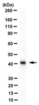
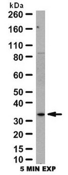
Evaluated by Western Blotting in HeLa nuclear extract.
Western Blotting Analysis: 0.5 µg/mL of this antibody detected Galectin-9 in 10 µg of HeLa nuclear extract.
Western Blotting Analysis: 0.5 µg/mL of this antibody detected Galectin-9 in 10 µg of HeLa nuclear extract.
표적 설명
~38 kDa observed
물리적 형태
Format: Purified
기타 정보
Concentration: Please refer to lot specific datasheet.
적합한 제품을 찾을 수 없으신가요?
당사의 제품 선택기 도구.을(를) 시도해 보세요.
Storage Class Code
12 - Non Combustible Liquids
WGK
WGK 2
Flash Point (°F)
Not applicable
Flash Point (°C)
Not applicable
시험 성적서(COA)
제품의 로트/배치 번호를 입력하여 시험 성적서(COA)을 검색하십시오. 로트 및 배치 번호는 제품 라벨에 있는 ‘로트’ 또는 ‘배치’라는 용어 뒤에서 찾을 수 있습니다.
A novel monoclonal antibody for detection of galectin-9 in tissue sections: application to human tissues infected by oncogenic viruses.
Barjon, C; Niki, T; Verillaud, B; Opolon, P; Bedossa, P; Hirashima, M; Blanchin et al.
Infectious Agents and Cancer null
자사의 과학자팀은 생명 과학, 재료 과학, 화학 합성, 크로마토그래피, 분석 및 기타 많은 영역을 포함한 모든 과학 분야에 경험이 있습니다..
고객지원팀으로 연락바랍니다.