추천 제품
제품명
Anti-beta-Actin Antibody, clone 4C2, clone 4C2, from mouse
생물학적 소스
mouse
Quality Level
항체 형태
purified immunoglobulin
항체 생산 유형
primary antibodies
클론
4C2, monoclonal
종 반응성
chicken, human, mouse, rat
종 반응성(상동성에 의해 예측)
mammals (based on 100% sequence homology)
기술
immunocytochemistry: suitable
western blot: suitable
동형
IgG1κ
NCBI 수납 번호
UniProt 수납 번호
배송 상태
wet ice
타겟 번역 후 변형
unmodified
유전자 정보
human ... ACTB(60)
일반 설명
Actin, cytoplasmic 1 (UniProt P60709; also known as Beta-actin) is encoded by the ACTB gene (Gene ID 60) in human. Actins are globular multi-functional proteins that serve as the basic building blocks of cytoskeletal microfilaments and are among the most conserved eukaryotic proteins. Six actin types exist, skeletal muscle alpha-actin is encoded by the ACTA1 gene, smooth muscle alpha-actin by the ACTA2 gene, cytoplasmic beta-actin by the ACTB gene, cardiac muscle alpha-actin by the ACTC1 gene, cytoplasmic gamma-actin by the ACTG1 gene, and smooth muscle gamma-actin by the ACTG2 (a.k.a. ACTA3) gene. Although actins show >90% overall sequence homology, isoforms do show spatial, temporal, and tissue-specific expression patterns and only 50-60% homology is found in their 18 N-terminal residues. Cytoplasmic β and γ-actins, are thought to be present in all cells, while the other four actin isoforms are typically found in specific adult muscle tissue types. Actins exist in a variety of structural states, depending on the specific ionic conditions or the interaction with ligand proteins. The oligomeric and polymeric forms that actin molecules assume are dependent on the distinct conformations they adopt.
특이성
Clone 4C2 detected BSA conjugated with beta-actin N-terminal peptide, but not BSA conjugated with N-terminal peptides derived from the 5 other actin types (Dugina, V., et al. (2009). J. Cell Sci. 122(Pt 16):2980-2988).
면역원
KLH-conjugated linear peptide corresponding to the N-terminal sequence of human beta-Actin.
애플리케이션
This Anti-beta-Actin Antibody, clone 4C2 is validated for use in Western Blotting, Immunocytochemistry for the detection of beta-Actin.
Western Blotting Analysis: 0.5 µg/mL from a representative lot detected beta-Actin in 10 µg of NIH/3T3 cell lysate.
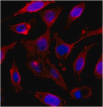
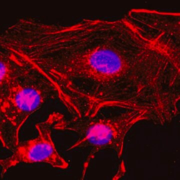
Immunocytochemistry Analysis: 5.0 µg/mL from a representative lot detected beta-Actin in HUVECs, A431 and HeLa cells.
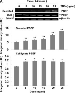
Western Blotting Analysis: A representative lot detected downregulated beta-actin levels in stimulated (by A23187, TRAP-6, TNF, LPS, or IFN-γ) human cerebral microvascular endothelial D3 cells (hCMEC/D3) and their microparticles (MPs) when compared with unstimulated hCMEC/D3 and their MPs (Latham, S.L., et al. (2013). FASEB J. 27(2):672-683).
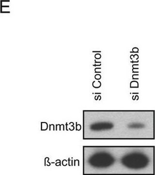
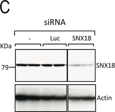
Western Blotting Analysis: A representative lot detected siRNA-mediated downregulation of beta-actin in A549 human lung carcinoma cells (Miazza, V., et al. (2011). Virology. 410(1):7-16).
Western Blotting Analysis: A representative lot detected beta-actin, but not cytoplasmic gamma-actin separated by 2-D gel electrophoresis of purified chicken gizzard actins or total protein extracts from human subcutaneous fibroblasts (HSCFs), canine MDCK cells, and rat aorta tissue (Dugina, V., et al. (2009). J. Cell Sci. 122(Pt 16):2980-2988).
Western Blotting Analysis: A representative lot detected BSA conjugated with beta-actin N-terminal peptide, but not BSA conjugated with N-terminal peptides derived from the other 5 actin types (Dugina, V., et al. (2009). J. Cell Sci. 122(Pt 16):2980-2988).
Immunocytochemistry Analysis: A representative lot detected TNF-stimulated localization of β-actin into thick, intensely staining stress fibers prominent at the basal surface of of human cerebral microvascular endothelial D3 cells (hCMEC/D3). Rho kinase inhibitor Y-27632 (Cat. No. 688000) treatment suppressed TNF-induced β-actin stress fiber formation (Latham, S.L., et al. (2013). FASEB J. 27(2):672-683).
Immunocytochemistry Analysis: A representative lot detected a drastic subcellular redistribution of beta-actin following Sendai virus infection of polarized Madin-Darby canine kidney (MDCK) epithelial cells by fluorescent immunocytochemistry staining of paraformaldehyde-fixed, methanol-treated cells (Miazza, V., et al. (2011). Virology. 410(1):7-16).
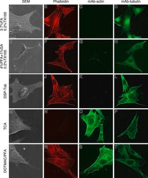
Immunocytochemistry Analysis: A representative lot detected beta-actin subcellular localization distinct from that of cytoplasmic gamma-actin in both spreading and stationary cells by fluorescent immunocytochemistry, using paraformaldehyde-fixed, methanol-treated HSCF human subcutaneous fibroblasts, HaCaT human keratinocytes, WI38 human embryonic fibroblasts,and Madin-Darby canine kidney (MDCK) cells (Dugina, V., et al. (2009). J. Cell Sci. 122(Pt 16):2980-2988).
품질
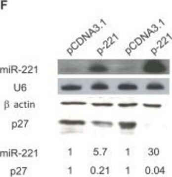
Evaluated by Western Blotting in HeLa cell lysate.
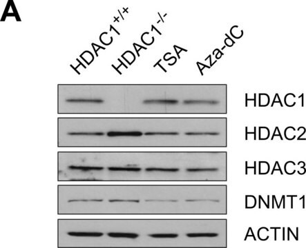
Western Blotting Analysis: 0.5 µg/mL of this antibody detected beta-Actin in 10 µg of HeLa cell lysate.
Western Blotting Analysis: 0.5 µg/mL of this antibody detected beta-Actin in 10 µg of HeLa cell lysate.
표적 설명
~39-45 kDa observed. Uncharacterized band(s) may appear in some lysates.
물리적 형태
Format: Purified
기타 정보
Concentration: Please refer to lot specific datasheet.
적합한 제품을 찾을 수 없으신가요?
당사의 제품 선택기 도구.을(를) 시도해 보세요.
Storage Class Code
12 - Non Combustible Liquids
WGK
WGK 1
Flash Point (°F)
Not applicable
Flash Point (°C)
Not applicable
시험 성적서(COA)
제품의 로트/배치 번호를 입력하여 시험 성적서(COA)을 검색하십시오. 로트 및 배치 번호는 제품 라벨에 있는 ‘로트’ 또는 ‘배치’라는 용어 뒤에서 찾을 수 있습니다.
이미 열람한 고객
Venu Venkatarame Gowda Saralamma et al.
Oncology reports, 44(3), 939-958 (2020-07-25)
Scutellarein (SCU), a flavone that belongs to the flavonoid family and abundantly present in Scutellaria baicalensis a flowering plant in the family Lamiaceae, has been reported to exhibit anticancer effects in several cancer cell lines including gastric cancer (GC). Although
Yan Lin et al.
Frontiers in veterinary science, 9, 904653-904653 (2022-08-02)
Sperm and seminal plasma are rich in leucine, and leucine can promote the protein synthesis. This property makes it an interesting amino acid to increase sperm quality of human and livestock spermatogenesis. The goal of this study was to explore
Rouven Schoppmeyer et al.
Cell reports, 38(3), 110243-110243 (2022-01-20)
Understanding how cytotoxic T lymphocytes (CTLs) efficiently leave the circulation to target cancer cells or contribute to inflammation is of high medical interest. Here, we demonstrate that human central memory CTLs cross the endothelium in a predominantly paracellular fashion, whereas
Rujun Chen et al.
Molecular medicine reports, 24(5) (2021-09-16)
Placenta‑specific protein 1 (PLAC1) is inversely associated with survival in several types of cancer. However, whether PLAC1 is involved in the progression of cervical cancer (CC) remains to be elucidated. Therefore, the present study aimed to evaluate the prognostic role of PLAC1 in
Xiaohu Huang et al.
Cancers, 13(4) (2021-02-13)
MYC and HIF1α are among the most important oncoproteins whose pharmacologic inhibition has been challenging for the diverse mechanisms driving their abnormal expression and because of the challenge in blocking protein-DNA interactions. Surprisingly, we found that MYC and HIF1α proteins
자사의 과학자팀은 생명 과학, 재료 과학, 화학 합성, 크로마토그래피, 분석 및 기타 많은 영역을 포함한 모든 과학 분야에 경험이 있습니다..
고객지원팀으로 연락바랍니다.