추천 제품
생물학적 소스
rat
Quality Level
결합
unconjugated
항체 형태
purified antibody
항체 생산 유형
primary antibodies
클론
B6A6, monoclonal
정제법
using protein G
종 반응성
mouse
포장
antibody small pack of 100 μg
기술
flow cytometry: suitable
immunohistochemistry (formalin-fixed, paraffin-embedded sections): suitable
동형
IgG2aκ
에피토프 서열
Unknown
단백질 ID 수납 번호
UniProt 수납 번호
배송 상태
ambient
타겟 번역 후 변형
unmodified
일반 설명
The small intestine consists of continuous villi and crypts. The crypt is mainly a proliferative compartment and is maintained by multipotent stem cells whereas the villus represents the differentiated compartment, and it receives cells from multiple crypts. The functional compartment of the epithelium contains differentiated cells that populate the villi and can be categorized based on their function: enterocytes that function to absorb nutrients, goblet cells that secrete a protective mucus barrier, and enteroendocrine cells that release gastrointestinal hormones. Intestinal crypts contain intestinal stem cells (ISC), which are multipotent adult stem cells that reside in the base of crypts. They continuously self-renew by dividing and differentiate into the specialized cells of the intestinal epithelium, which renews throughout life. ISCs located either at the crypt base interspersed between the Paneth cells can be categorized into active-cycling (Lgr5+ cells) or those located near the þ4 position in the crypt as slow-cycling Bmi-1+ cells. Paneth cells that are responsible for secreting antibacterial peptides reside at the base of the proliferative compartment. As ISCs begin to differentiate, they migrate toward the lumen and are eventually shed, either from the tip of the intestinal villi or from the surface of the colonic epithelium. Villus represents the differentiated compartment of intestinal cells. Clone B6A6 recognizes most villous cells except goblet cells. (Ref.: Smith, NR., et al. (2018). Cell. Mol. Gastroenterol. Hepatol. 6(1);79-96; Wang, F., et al. (2013). Gastroenterology. 145(2); 383-395; Umar, S. (2010). Curr. Gastroenterol. Rep. 12(5); 340-348).
특이성
Clone B6A6 is a rat monoclonal antibody that detects intestinal villus cells.
면역원
Undifferentiated Mouse intestinal epithelial cells. Rats were preimmunized with isolations of differentiated mouse intestinal epithelial cells and then treated with cyclophosphamide (i.p.) to eliminate B lymphocytes reacting against these antigens. Subsequent immunization was with crypt-based single cells.
애플리케이션
Quality Control Testing
Isotype testing: Identity Confirmation by Isotyping Test.
Isotyping Analysis: The identity of this monoclonal antibody is confirmed by isotyping test to be mouse IgG2a .
Tested Applications
Flow Cytometry Analysis: A representative lot detected Intestinal Villus in Lgr5-GFP mouse intestinal cells ( Data courtesy of Prof. Melissa Wong, Ph.D., Oregon Health & Science University, Portland, Oregon USA).
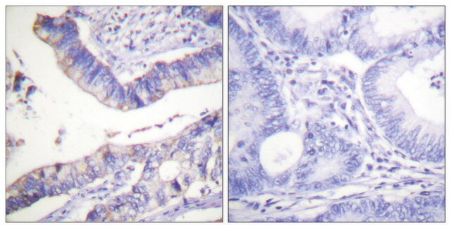
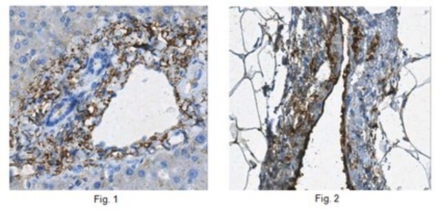
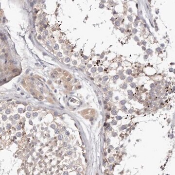
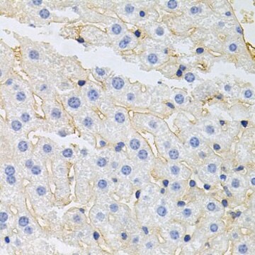
Immunohistochemistry Applications: A representative lot detected Intestinal Villus cells in Immunohistochemistry applications (Wang, F., et al. (2013). Gastroenterology. 145(2): 383-95.e1-21).
Flow Cytometry Analysis: A representative lot detected Intestinal Villus in Flow Cytometry applications (Wang, F., et al. (2013). Gastroenterology. 145(2): 383-95.e1-21; Smith, N.R., et al. (2017). Cell Mol Gastroenterol Hepatol. 3(3): 389-409; Yan, K.S., et al. (2017). Cell Stem Cell. 21(1):7 8-90.e6; Smith, N.R., et al. (2018). Cell Mol Gastroenterol Hepatol. 6(1): 79-96).
Note: Actual optimal working dilutions must be determined by end user as specimens, and experimental conditions may vary with the end user
Anti-Intestinal Villus clone B6A6, Cat. No. MABS2233, is a rat monoclonal antibody that detects Intestinal Villus cells and is tested for use in and Flow Cytometry and Immunohistochemistry.
물리적 형태
Purified rat monoclonal antibody IgG2a in buffer containing 0.1 M Tris-Glycine (pH 7.4), 150 mM NaCl with 0.05% sodium azide.
저장 및 안정성
Recommended storage: +2°C to +8°C.
기타 정보
Concentration: Please refer to the Certificate of Analysis for the lot-specific concentration.
면책조항
Unless otherwise stated in our catalog or other company documentation accompanying the product(s), our products are intended for research use only and are not to be used for any other purpose, which includes but is not limited to, unauthorized commercial uses, in vitro diagnostic uses, ex vivo or in vivo therapeutic uses or any type of consumption or application to humans or animals.
적합한 제품을 찾을 수 없으신가요?
당사의 제품 선택기 도구.을(를) 시도해 보세요.
Storage Class Code
12 - Non Combustible Liquids
WGK
WGK 1
시험 성적서(COA)
제품의 로트/배치 번호를 입력하여 시험 성적서(COA)을 검색하십시오. 로트 및 배치 번호는 제품 라벨에 있는 ‘로트’ 또는 ‘배치’라는 용어 뒤에서 찾을 수 있습니다.
자사의 과학자팀은 생명 과학, 재료 과학, 화학 합성, 크로마토그래피, 분석 및 기타 많은 영역을 포함한 모든 과학 분야에 경험이 있습니다..
고객지원팀으로 연락바랍니다.