
MABS1726
Anti-MPK-3 (DUSP6) Antibody, clone 5H6.1
clone 5H6.1, from mouse
동의어(들):
Dual specificity protein phosphatase 6, Dual specificity protein phosphatase PYST1, MAP kinase phosphatase 3, Mitogen-activated protein kinase phosphatase 3, MKP-3
로그인조직 및 계약 가격 보기
모든 사진(2)
About This Item
UNSPSC 코드:
12352203
eCl@ss:
32160702
추천 제품


생물학적 소스
mouse
Quality Level
항체 형태
purified antibody
항체 생산 유형
primary antibodies
클론
5H6.1, monoclonal
종 반응성
human
기술
immunohistochemistry: suitable (paraffin)
western blot: suitable
동형
IgG2bκ
NCBI 수납 번호
UniProt 수납 번호
배송 상태
ambient
타겟 번역 후 변형
unmodified
유전자 정보
human ... DUSP6(1848)
일반 설명
Dual specificity protein phosphatase 6 (EC 3.1.3.16; EC 3.1.3.48; UniProt Q16828; also known as Dual specificity protein phosphatase PYST1, MAP kinase phosphatase 3, Mitogen-activated protein kinase phosphatase 3, MKP-3) is encoded by the DUSP6 (also known as HH19, MKP3, PYST1) gene (Gene ID 1848) in human. MPK-3 (DUSP6) belongs to the family of dual-specificity phosphatases (DUSPs) that mediate dephosphorylation of MAPKs on threonine/serine and tyrosine residues. DUSP6 is upregulated in HER2+ carcinomas and is among several DUSPs thought to be involved in breast cancer metastasis. DUSP1, DUSP4, and DUSP6 are induced during epithelial-to-mesenchymal transition (EMT) and are involved in forming and maintaining breast cancer stem cells (CSCs), DUSP1 knockdown reduces CSC formation, while DUSP4 or DUSP6 knockdown enhances CSC formation. PKC CK2alpha is reported to phosphorylate DUSP6 and increase its dephosphatase activity. ERK1/2, on the other hand, can phosphorylate DUSP6 to promote its proteosomal degradation.
특이성
Clone 5H6.1 targets an epitope within the rhodanese domain present in both spliced isoforms of human MPK-3 (DUSP6) reported by UniProt (Q16828).
면역원
GST-tagged recombinant human MPK-3 (DUSP6) rhodanese domain fragment.
애플리케이션
Detect DUSP6 using this mouse monoclonal Anti-MPK-3 (DUSP6), clone 5H6.1 Antibody, Cat. No. MABS1726, validated for use in Immunohistochemistry (Paraffin) and Western Blotting.
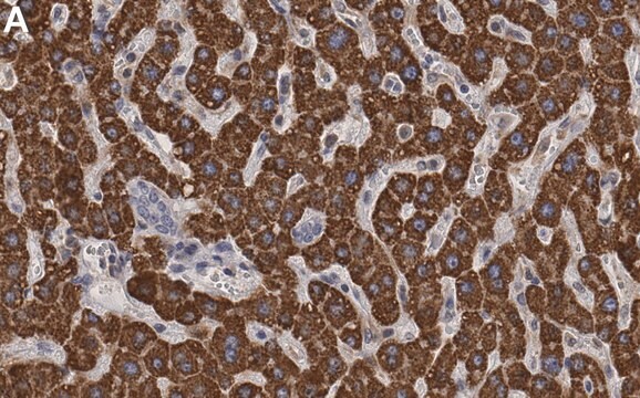
Immunohistochemistry Analysis: A 1:1,000 dilution from a representative lot detected MPK-3 (DUSP6) in human liver, pancreas, and tonsil tissue sections.
Research Category
Signaling
Signaling
품질
Evaluated by Western Blotting in HepG2 cell lysate.
Western Blotting Analysis: 0.5 µg/mL of this antibody detected MPK-3 (DUSP6) in 10 µg of HepG2 cell lysate.
Western Blotting Analysis: 0.5 µg/mL of this antibody detected MPK-3 (DUSP6) in 10 µg of HepG2 cell lysate.
표적 설명
~42 kDa observed. 42.32 kDa (isoform 1) and 26.47 kDa (isoform 2; DUSP6-ALT) calculated. Uncharacterized bands may be observed in some lysate(s).
물리적 형태
Format: Purified
Protein G purified.
Purified mouse IgG2bκ in buffer containing 0.1 M Tris-Glycine (pH 7.4), 150 mM NaCl with 0.05% sodium azide.
저장 및 안정성
Stable for 1 year at 2-8°C from date of receipt.
기타 정보
Concentration: Please refer to lot specific datasheet.
면책조항
Unless otherwise stated in our catalog or other company documentation accompanying the product(s), our products are intended for research use only and are not to be used for any other purpose, which includes but is not limited to, unauthorized commercial uses, in vitro diagnostic uses, ex vivo or in vivo therapeutic uses or any type of consumption or application to humans or animals.
적합한 제품을 찾을 수 없으신가요?
당사의 제품 선택기 도구.을(를) 시도해 보세요.
Storage Class Code
12 - Non Combustible Liquids
WGK
WGK 1
Flash Point (°F)
Not applicable
Flash Point (°C)
Not applicable
시험 성적서(COA)
제품의 로트/배치 번호를 입력하여 시험 성적서(COA)을 검색하십시오. 로트 및 배치 번호는 제품 라벨에 있는 ‘로트’ 또는 ‘배치’라는 용어 뒤에서 찾을 수 있습니다.
자사의 과학자팀은 생명 과학, 재료 과학, 화학 합성, 크로마토그래피, 분석 및 기타 많은 영역을 포함한 모든 과학 분야에 경험이 있습니다..
고객지원팀으로 연락바랍니다.