추천 제품
생물학적 소스
mouse
Quality Level
항체 형태
purified antibody
항체 생산 유형
primary antibodies
클론
IgG-A9, monoclonal
종 반응성
human, rat, hamster
기술
immunocytochemistry: suitable
immunoprecipitation (IP): suitable
western blot: suitable
동형
IgG1κ
UniProt 수납 번호
배송 상태
ambient
타겟 번역 후 변형
unmodified
유전자 정보
human ... HMGCR(3156)
일반 설명
3-hydroxy-3-methylglutaryl-coenzyme A reductase (EC 1.1.1.34; UniProt P00347; also known as HMG-CoA reductase) is encoded by the HMGCR gene (Gene ID 100756363) in Cricetulus griseus (Chinese hamster). HMG-CoA reductase catalyzes the conversion of HMG-CoA to mevalonic acid, the rate-limiting enzyme of the mevalonate-isoprenoid biosynthesis (MIB) pathway responsible for the production of cholesterol and other isoprenoids, as well as farnesyl pyrophosphate (FPP) required for protein prenylation. HMG-CoA reductase is suppressed by cholesterol derived from the internalization and degradation of low density lipoprotein (LDL) via the LDL receptor as well as oxidized cholesterol species. Competitive inhibitors against HMG-CoA reductase induce LDL receptors expression in the liver, which in turn increases the catabolism of plasma LDL and lowers the plasma concentration of cholesterol. HMG-CoA reductase is a multitransmembrane ER protein with an N-terminal sterol-sensing domain (SSD; a.a. 61-218) and a C-terminal catalytic domain (a.a. 450-888) whose activity is inhibited upon SSD binding by cholesterol. Numerous studies have implicated the involvement of HMG-CoA reductase and mevalonate pathway in carcinogenesis.
특이성
Clone IgG-A9 targets an epitope within HMC-CoA reductase catalytic domain (a.a. 450 887).
면역원
Membrane preapration from compactin-adapted CHO cells (Liscum, L., et al. (1983). J. Biol. Chem. 258(13) 8450-8455).
애플리케이션
Detect HMG CoA Reductase using this mouse monoclonal Anti-HMG-CoA Reductase, clone IgG-A9, Cat. No. MABS1233, validated for use in Immunocytochemistry, Immunoprecipitation, and Western Blotting.
Research Category
Signaling
Signaling
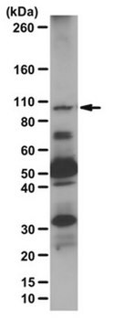
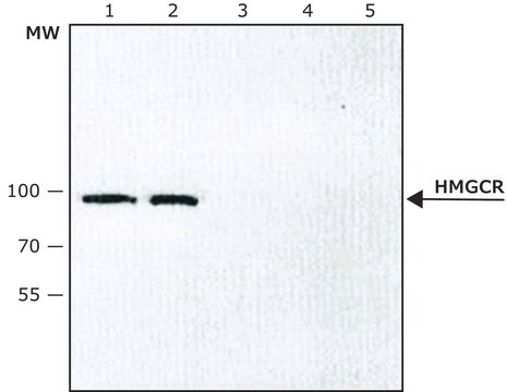
Western Blotting Analysis: A 1:100 dilution from a representative lot detected in 15 µg of membrane extract from CHO7 cells treated overnight with 0.01 mM compactin (Courtesy of Linda Donnelly, Department of Molecular Genetics, UT Southwestern Medical Center, Dallas, TX, USA).
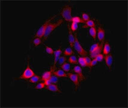
Immunocytochemistry Analysis: A representative lot detected HMG CoA reductase-positive ER membrane structures by indirect immunofluorescence staining of paraformaldehyde-fixed, 0.1% Triton X-100-permeabilized CHO-K1 cells cultured with compactin, mevalonate, and MG-132, in the presence or absence of 25-hydroxycholesterol (25-HC) (Hartman, I.Z., et al. (2010). J. Biol. Chem. 285(25):19288-19298).
Immunoprecipitation Analysis: A representative lot immunodepleted HMC-CoA reductase activity from compactin-adapted CHO cell (UT-1) extract (Liscum, L., et al. (1983). J. Biol. Chem. 258(13) 8450-8455).
Western Blotting Analysis: A representative lot detected 25-hydroxycholesterol (25-HC) treatment-induced HMC-CoA reductase dislocation from ER membrane to the cytosol and lipid droplet in CHO-K1 cells cultured with lipoprotein-deficient serum in the presence of compactin, MG-132, with or without mevalonate. Cellular VCP knockdown prevented sterol-induced HMC-CoA reductase cytosolic, but not lipid droplet, translocation (Hartman, I.Z., et al. (2010). J. Biol. Chem. 285(25):19288-19298).
Western Blotting Analysis: A representative lot detected 25-hydroxycholesterol (25-HC) treatment-induced ubiquitination of HMC-CoA reductase in SV-589 human fibroblasts cultured with compactin and mevalonate in the presence of proteasome inhibitor MG-132. Cellular knockdown of gp78A, but not gp78B or Hrd1, reduced 25-HC-induced HMC-CoA reductase ubiquitination (Song, B.L., et al. (2005). Mol. Cell. 19(6):829-840).
Western Blotting Analysis: A representative lot detected 25-hydroxycholesterol (25-HC) treatment-induced degradation of HMC-CoA reductase in SV-589 human fibroblasts cultured with compactin and mevalonate. Cellular knockdown of gp78A, VCP, or Insig-1/-2 suppressed sterol-induced HMC-CoA reductase degradation (Song, B.L., et al. (2005). Mol. Cell. 19(6):829-840).
Western Blotting Analysis: A representative lot detected HMC-CoA reductase in both membrane and nuclear fractions from HEK-293S cells cultured with compactin and mevalonate. Additional treatment with 25-hydroxycholesterol (25-HC) resulted in HMC-CoA reductase degradation (Sever, N., et al. (2003). Mol. Cell. 11(1):25-33).
Western Blotting Analysis: A representative lot detected an upregulated HMC-CoA reductase expression in compactin-adapted CHO cells (UT-1), as well as a loss of HMC-CoA reductase expression in UT-1 cells cultured in the presence of LDL (Liscum, L., et al. (1983). J. Biol. Chem. 258(13) 8450-8455).
Western Blotting Analysis: A representative lot detected a greater HMC-CoA reductase upregulation in liver microsome preparation from rats on diet supplemented with cholestyramine and mevinolin than with cholestyramine alone, while supplementation with cholesterol in addition to cholestyramine and mevinolin downregulated liver HMC-CoA reductase (Liscum, L., et al. (1983). J. Biol. Chem. 258(13) 8450-8455).
품질
Identity Confirmation by Isotyping Test.
Isotyping Analysis: The identity of this monoclonal antibody is confirmed by isotyping test to be mouse IgG1 .
Isotyping Analysis: The identity of this monoclonal antibody is confirmed by isotyping test to be mouse IgG1 .
표적 설명
~92 kDa observed. 97.08 kDa (hamster), 97.48/92.02/99.75 kDa (human isoform 1/2/3 (HMGCR-1b)), 96.69 kDa (rat) calculated.. Uncharacterized bands may be observed in some lysate(s).
물리적 형태
Format: Purified
Protein G purified.
Purified mouse IgG1 in buffer containing 0.1 M Tris-Glycine (pH 7.4), 150 mM NaCl with 0.05% sodium azide.
저장 및 안정성
Stable for 1 year at 2-8°C from date of receipt.
기타 정보
Concentration: Please refer to lot specific datasheet.
면책조항
Unless otherwise stated in our catalog or other company documentation accompanying the product(s), our products are intended for research use only and are not to be used for any other purpose, which includes but is not limited to, unauthorized commercial uses, in vitro diagnostic uses, ex vivo or in vivo therapeutic uses or any type of consumption or application to humans or animals.
적합한 제품을 찾을 수 없으신가요?
당사의 제품 선택기 도구.을(를) 시도해 보세요.
Storage Class Code
12 - Non Combustible Liquids
WGK
WGK 1
Flash Point (°F)
Not applicable
Flash Point (°C)
Not applicable
시험 성적서(COA)
제품의 로트/배치 번호를 입력하여 시험 성적서(COA)을 검색하십시오. 로트 및 배치 번호는 제품 라벨에 있는 ‘로트’ 또는 ‘배치’라는 용어 뒤에서 찾을 수 있습니다.
자사의 과학자팀은 생명 과학, 재료 과학, 화학 합성, 크로마토그래피, 분석 및 기타 많은 영역을 포함한 모든 과학 분야에 경험이 있습니다..
고객지원팀으로 연락바랍니다.