추천 제품
생물학적 소스
mouse
Quality Level
항체 형태
purified antibody
항체 생산 유형
primary antibodies
클론
10.22, monoclonal
종 반응성
rat, mouse, human, fish, bovine
기술
immunofluorescence: suitable
immunohistochemistry: suitable (paraffin)
immunoprecipitation (IP): suitable
western blot: suitable
동형
IgG1
NCBI 수납 번호
UniProt 수납 번호
배송 상태
wet ice
타겟 번역 후 변형
unmodified
유전자 정보
human ... SYN1(6853)
일반 설명
Synapsin-1 (UniProt P17599; also known as Synapsin I) is encoded by the SYN1 gene (Gene ID 281510) in bovine species. The synapins constitute a family of abundant neuronal phosphoproteins that regulate SV trafficking and neurotransmitter release at the pre-synaptic terminal. Three genes exisit in mammals, encoding altogether 11 synapsin members by alternative splicings (Synapsin Ia & Ib by SYN1, Synapsin IIa & IIb by SYN2, Synapsin IIIa through IIIf by SYN3), Syn III is the most precociously expressed isoform that has a role in the early phases of neural development and is downregulated in mature neurons. On the other hand, Syn I and Syn II are expressed at low levels at birth and their expressions progressively increase during synaptogenesis to reach a stable plateau at 1–2 months of life. The NH2-terminal region is divided into A, B, and C domains and is highly conserved among synapsin isoforms. Synapsins are regulated by postransloational phosphorylations. Domain A contains PKA and CaMKI/IV phosphorylation sites, domain B contains MAPK/Erk phosphorylation sites, and domain C is phosphorylated by Src. The C-terminal region contains spliced domains and diverge among synapsin isoforms (domain D in Syn Ia and Ib, domain G in Syn IIa and IIb, domain H in Syn IIa, and domain J in Syn IIIa), although they all bear proline-rich regions binding to several SH3-containing proteins and additional phosphorylation sites for CaMKII, MAPK/Erk, and cdk1/5.
특이성
Clone 10.22 reacts with both synapsin-1 spliced isoforms (Ia and Ib), but not synapsin-2 spliced isoform IIa or IIb (Vaccaro, P., et al. (1997). Brain Res. Mol. Brain Res. 52(1):1-16).
면역원
Epitope: Pro-rich domain D.
Purified bovine brain synapsin-1.
애플리케이션
Immunohistochemistry Analysis: A 1:50 dilution from a representative lot detected Synapsin-1 in human, mouse, and rat cerebral cortex tissues.
Western Blotting Analysis: A representative lot detected synapsin Ia/Ib in mouse cortical neuron lysates (Medrihan, L., et al. (2013). Nat. Commun. 4:1512).
Western Blotting Analysis: A representative lot detected purified bovine brain synapsin Ia/Ib (Messa, M., et al. (2010). J. Cell Sci. 123(13):2256-2265).
Western Blotting Analysis: A representative lot detected synapsin-1 in Torpedo (electric ray) synaptosomal preparations and in GST-cyclophilin B pull-downs (Lane-Guermonprez, L., et al. (2005). J. Neurochem. 93(6):1401-1411).
Western Blotting Analysis: A representative lot detected synapsin-1 in the same rat brain subcellular fractions as cyclophilin B (Lane-Guermonprez, L., et al. (2005). J. Neurochem. 93(6):1401-1411).
Western Blotting Analysis: A representative lot detected synapsin Ia/Ib, but not IIa/IIb, in rat brain post-nuclear supernatants (Vaccaro, P., et al. (1997). Brain Res. Mol. Brain Res. 52(1):1-16).
Immunoprecipitation Analysis: A representative lot immunoprecipitated synapsin Ia/Ib, but not IIa/IIb, from rat brain synaptosomal preparations using protein G beads with pre-bound rabbit anti-mouse IgG (Vaccaro, P., et al. (1997). Brain Res. Mol. Brain Res. 52(1):1-16).
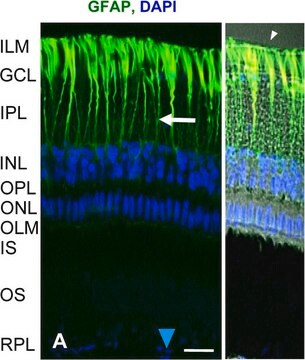
Immunofluorescence Analysis: Clone 10.22 ascites fluid was employed to localize synapsin-1 immunoreactivity within retinal inner plexiform layer (IPL) of floating or whole-mount rat retinas (Mandell, J.W., et al. (1992). J. Neurosci. 12(5):1736-1749).
Note: Clone 10.22 does not bind significantly to protein G. For immunoprecipitation application, pre-coat protein G beads with an anti-mouse IgG antibody is recommended (Vaccaro, P., et al. (1997). Brain Res. Mol. Brain Res. 52(1):1-16).
Research Category
Neuroscience
Neuroscience
Research Sub Category
Developmental Neuroscience
Developmental Neuroscience
This Anti-Synapsin-1 Antibody, clone 10.22 is validated for use in Western Blotting, Immunohistochemistry (Paraffin), Immunoprecipitation, Immunofluorescence for the detection of Synapsin-1.
품질
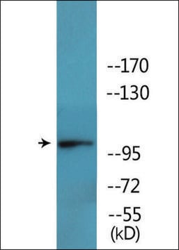
Evaluated by Western Blotting in rat brain cytosol tissue lysate.
Western Blotting Analysis: 0.5 µg/mL of this antibody detected Synapsin-1 in 10 µg of rat brain cytosol tissue lysate.
Western Blotting Analysis: 0.5 µg/mL of this antibody detected Synapsin-1 in 10 µg of rat brain cytosol tissue lysate.
표적 설명
~75 kDa observed.
물리적 형태
Format: Purified
Protein G Purified
Purified mouse monoclonal IgG1 antibody in buffer containing 0.1 M Tris-Glycine (pH 7.4), 150 mM NaCl with 0.05% sodium azide.
저장 및 안정성
Stable for 1 year at 2-8°C from date of receipt.
기타 정보
Concentration: Please refer to lot specific datasheet.
면책조항
Unless otherwise stated in our catalog or other company documentation accompanying the product(s), our products are intended for research use only and are not to be used for any other purpose, which includes but is not limited to, unauthorized commercial uses, in vitro diagnostic uses, ex vivo or in vivo therapeutic uses or any type of consumption or application to humans or animals.
적합한 제품을 찾을 수 없으신가요?
당사의 제품 선택기 도구.을(를) 시도해 보세요.
Storage Class Code
12 - Non Combustible Liquids
WGK
WGK 1
Flash Point (°F)
Not applicable
Flash Point (°C)
Not applicable
시험 성적서(COA)
제품의 로트/배치 번호를 입력하여 시험 성적서(COA)을 검색하십시오. 로트 및 배치 번호는 제품 라벨에 있는 ‘로트’ 또는 ‘배치’라는 용어 뒤에서 찾을 수 있습니다.
Kanzo Suzuki et al.
Cell reports, 37(5), 109918-109918 (2021-11-04)
Ketamine is a noncompetitive glutamatergic N-methyl-d-aspartate receptor (NMDAR) antagonist that exerts rapid antidepressant effects. Preclinical studies identify eukaryotic elongation factor 2 kinase (eEF2K) signaling as essential for the rapid antidepressant action of ketamine. Here, we combine genetic, electrophysiological, and pharmacological
자사의 과학자팀은 생명 과학, 재료 과학, 화학 합성, 크로마토그래피, 분석 및 기타 많은 영역을 포함한 모든 과학 분야에 경험이 있습니다..
고객지원팀으로 연락바랍니다.

