
추천 제품

일반 설명
PAH, also known as Phenylalanine-4-hydroxylase , Phe-4-monooxygenase, and encoded by the gene name PAH, belongs to the biopterin-dependent aromatic amino acid hydroxylase family. Phenylalanine hydroxylase is the rate-limiting enzyme of the metabolic pathway that degrades excess phenylalanine. Phenylalanine hydroxylase (PheOH, alternatively PheH or PAH) is an enzyme that catalyzes the hydroxylation of the aromatic side-chain of phenylalanine to generate tyrosine. PheOH is one of three members of the pterin-dependent amino acid hydroxylases, a class of monooxygenase that uses tetrahydrobiopterin (BH4, a pteridine cofactor) and a non-heme iron for catalysis. During the reaction, molecular oxygen is heterolytically cleaved with sequential incorporation of one oxygen atom into BH4 and phenylalanine substrate. PAH has been associated with Phenylketonuria PKU, an autosomal recessive inborn error of phenylalanine metabolism, due to severe phenylalanine hydroxylase deficiency. Additioanlly, PAH has been associated with Non-phenylketonuria hyperphenylalaninemia (Non-PKU HPA), a mild form of phenylalanine hydroxylase deficiency characterized by phenylalanine levels persistently below 600 mumol, which allows normal intellectual and behavioral development without treatment. Finally, PAH may play a role in the Hyperphenylalaninemia (HPA), a mildest form of phenylalanine hydroxylase deficiency. PAH is broadly expressed, with greatest levels in skeletal muscle followed by heart, brain, pancreas and testis.
면역원
GST-tagged recombinant protein corresponding to human PAH.
애플리케이션
This Anti-PAH antibody is validated for use in WB, IH for the detection of PAH.
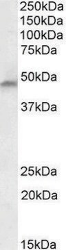
Western Blotting Analysis: 1.0 µg/mL from a representative lot detected PAH in 10 µg of human liver tissue lysate.
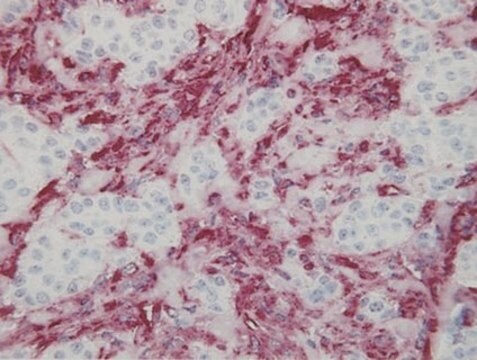
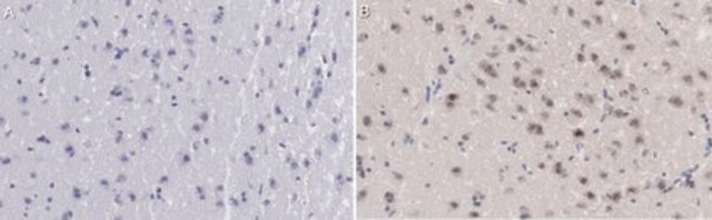
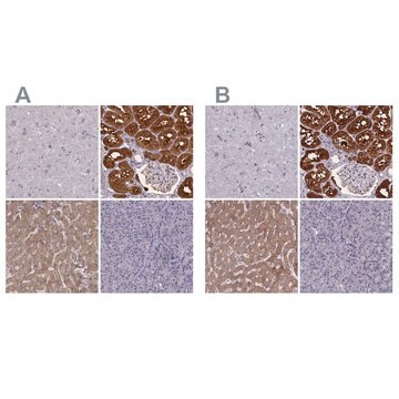
Immunohistochemistry Analysis: A 1:50-250 dilution from a representative lot detected PAH in human cerebral cortex and human liver tissue.
Immunohistochemistry Analysis: A 1:50-250 dilution from a representative lot detected PAH in human cerebral cortex and human liver tissue.
품질
Evaluated by Western Blotting in HepG2 cell lysate.
Western Blotting Analysis: 1.0 µg/mL of this antibody detected PAH in 10 µg of HepG2 cell lysate.
Western Blotting Analysis: 1.0 µg/mL of this antibody detected PAH in 10 µg of HepG2 cell lysate.
표적 설명
~52 kDa observed
물리적 형태
Format: Purified
기타 정보
Concentration: Please refer to lot specific datasheet.
적합한 제품을 찾을 수 없으신가요?
당사의 제품 선택기 도구.을(를) 시도해 보세요.
Storage Class Code
12 - Non Combustible Liquids
WGK
WGK 1
Flash Point (°F)
Not applicable
Flash Point (°C)
Not applicable
시험 성적서(COA)
제품의 로트/배치 번호를 입력하여 시험 성적서(COA)을 검색하십시오. 로트 및 배치 번호는 제품 라벨에 있는 ‘로트’ 또는 ‘배치’라는 용어 뒤에서 찾을 수 있습니다.
자사의 과학자팀은 생명 과학, 재료 과학, 화학 합성, 크로마토그래피, 분석 및 기타 많은 영역을 포함한 모든 과학 분야에 경험이 있습니다..
고객지원팀으로 연락바랍니다.