추천 제품
생물학적 소스
mouse
Quality Level
결합
unconjugated
항체 형태
purified antibody
항체 생산 유형
primary antibodies
클론
7G6, monoclonal
분자량
calculated mol wt 42.67 kDa
정제법
using protein G
종 반응성
human, monkey, bovine
포장
antibody small pack of 100 μg
기술
immunofluorescence: suitable
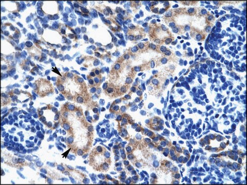
immunohistochemistry (formalin-fixed, paraffin-embedded sections): suitable
western blot: suitable
동형
IgG1κ
에피토프 서열
N-terminal
UniProt 수납 번호
배송 상태
2-8°C
타겟 번역 후 변형
unmodified
유전자 정보
mouse ... arr3> ARR3(102134564)
일반 설명
Arrestin-C (UniProt: A0A2K5U9A7; also known as Cone arrestin, Retinal cone arrestin-3) is encoded by the ARR3 gene (Gene ID: 102134564) in Monkey. Arrestins are a superfamily of multi-functional proteins that that regulate signaling and trafficking of the majority of G-protein-coupled receptors (GPCRs), as well as sub-cellular localization and activity of many other signaling proteins. Arrestin-C is predominantly found in inner and outer segments, and the inner plexiform regions of the retina. It is expressed in cone photoreceptors and pinealocytes and may contribute to the shut-off mechanisms associated with high acuity color vision. Arrestin-C is an elongated two-domain molecule with overall fold and key inter-domain interactions that hold the free protein in the basal conformation similar to the other subtypes. Several structural elements are reported to contribute to arrestin binding. The C-terminal acidic region serves a regulatory role in controlling arrestin binding selectivity toward the phosphorylated and activated form of a receptor. The basic N-terminal domain directly participates in receptor interaction and serves a regulatory role via intramolecular interaction with the C-terminal acidic region. Also, two centrally localized domains are directly involved in determining receptor binding specificity and selectivity. Clone 7G6 is a cone-specific marker that recognizes all cones in the adult primate retina. In fetal retina, although long and middle wavelength-sensitive cones recognized by clone 7G6, a subset of short wavelength-sensitive cones are shown to be delayed in their reactivity to this clone. (Ref.: Zhang, H., et al. (2003). Invest. Opthalmol. Vis. Sci. 44(7); 2858-2867; Wikler, KC., et al. (1997). J. Comp. Neurol. 377(4); 500-508).
특이성
Clone 7G6 is a mouse monoclonal antibody that detects cone arrestin (Arrestin-C). It targets an epitope within the N-terminal region.
면역원
Crude retinal extract from Monkey (Macaca fascicularis).
애플리케이션
Quality Control Testing
Isotype testing: Identity Confirmation by Isotyping Test.
Isotyping Analysis: The identity of this monoclonal antibody is confirmed by isotyping test to be mouse IgG1.
Tested Applications
Western Blotting Analysis: A representative lot detected Arrestin-C in Western Blotting applications (Zhang, H., et al. (2003). Invest Ophthalmol Vis Sci.;44(7):2858-67).
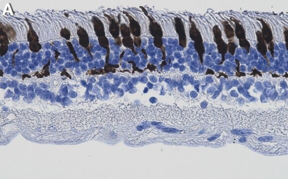
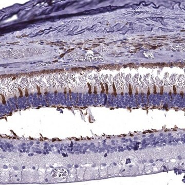
Immunohistochemistry Applications: A representative lot detected Arrestin-C in Immunohistochemistry applications (Wikler, K.C., et al. (1997). J Comp Neurol.;377(4):500-8; John, S.K., et al. (2000). Mol Vis.;6:204-15; Zhang, H., et al. (2003). Invest Ophthalmol Vis Sci.;44(7):2858-67; O′Brien, J.J., et al. (2012). J Neurosci.;32(13):4675-87).
Immunofluorescence Analysis: A 1:500 dilution from a representative lot detected Arrestin-C in frozen monkey retina tissues (Courtesy of Prof. Peter MacLeish, Ph.D., photo by Talib Saafir, Ph.D., Morehouse School of Medicine, Atlanta, GA, USA).
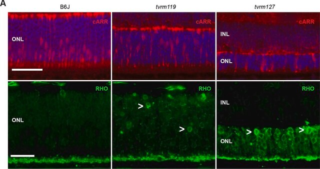
Immunofluorescence Analysis: A representative lot detected Arrestin-C in Immunofluorescence applications (Zhang, H., et al. (2003). Invest Ophthalmol Vis Sci.;44(7):2858-67).
Note: Actual optimal working dilutions must be determined by end user as specimens, and experimental conditions may vary with the end user
Anti-Arrestin-C, clone 7G6, Cat. No. MABN2636, is a mouse monoclonal antibody that detects Arrestin-C and is tested for use in Immunofluorescence, Immunohistochemistry, and Western Blotting.
물리적 형태
Purified mouse monoclonal antibody IgG1 in buffer containing 0.1 M Tris-Glycine (pH 7.4), 150 mM NaCl with 0.05% sodium azide.
저장 및 안정성
Recommend storage at +2°C to +8°C. For long term storage antibodies can be kept at -20°C. Avoid repeated freeze-thaws.
기타 정보
Concentration: Please refer to the Certificate of Analysis for the lot-specific concentration.
면책조항
Unless otherwise stated in our catalog or other company documentation accompanying the product(s), our products are intended for research use only and are not to be used for any other purpose, which includes but is not limited to, unauthorized commercial uses, in vitro diagnostic uses, ex vivo or in vivo therapeutic uses or any type of consumption or application to humans or animals.
적합한 제품을 찾을 수 없으신가요?
당사의 제품 선택기 도구.을(를) 시도해 보세요.
Storage Class Code
12 - Non Combustible Liquids
WGK
WGK 1
Flash Point (°F)
Not applicable
Flash Point (°C)
Not applicable
시험 성적서(COA)
제품의 로트/배치 번호를 입력하여 시험 성적서(COA)을 검색하십시오. 로트 및 배치 번호는 제품 라벨에 있는 ‘로트’ 또는 ‘배치’라는 용어 뒤에서 찾을 수 있습니다.
자사의 과학자팀은 생명 과학, 재료 과학, 화학 합성, 크로마토그래피, 분석 및 기타 많은 영역을 포함한 모든 과학 분야에 경험이 있습니다..
고객지원팀으로 연락바랍니다.