추천 제품
생물학적 소스
mouse
Quality Level
항체 형태
purified antibody
항체 생산 유형
primary antibodies
클론
KPSA1, monoclonal
종 반응성
bovine, human, mouse
포장
antibody small pack of 100
기술
direct ELISA: suitable
immunohistochemistry (formalin-fixed, paraffin-embedded sections): suitable
western blot: suitable
동형
IgG1κ
에피토프 서열
C-terminal
단백질 ID 수납 번호
UniProt 수납 번호
유전자 정보
human ... RPE65(6121)
특이성
Clone KPSA1 is a mouse monoclonal antibody that detects RPE65. It targets an epitope within 23 amino acid from the C-terminal region.
면역원
Recombinant full-length Human RPE65 expressed in Sf9 cells.
애플리케이션
Quality Control Testing
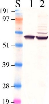
Evaluated by Western Blotting in Bovine retina microsomal preparation.
Western Blotting Analysis: A 1:10,000 dilution of this antibody detected RPE65 in Bovine retina microsomal preparation.
Tested Applications
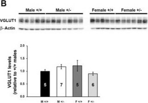
Western Blotting Analysis: A representative lot detected RPE65 in Western Blotting applications (Golczak, M., et al. (2010). J Biol Chem. 285(13):9667-9682; Banskota, S., et al. (2022). Cell. 185(2):250-265.e16).
Immunoaffinity Purification: A representative lot was used for purification of crossed-linked RPE65.(Golczak, M., et al. (2010). J Biol Chem. 285(13):9667-9682).
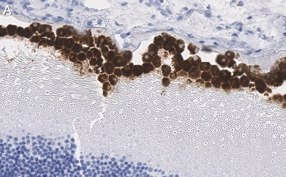
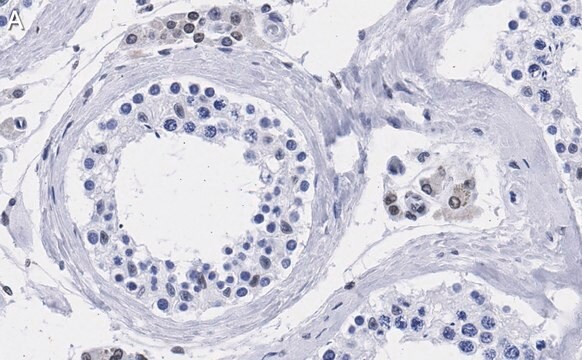
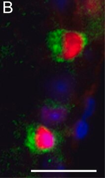
Immunohistochemistry Applications: A representative lot detected RPE65 in Immunohistochemistry applications (Amengual, J., et al. (2014). Hum Mol Genet. 23(20):5402-17).
ELISA Analysis: A representative lot detected RPE65 in ELISA applications (Golczak, M., et al. (2010). J Biol Chem. 285(13):9667-9682).
Note: Actual optimal working dilutions must be determined by end user as specimens, and experimental conditions may vary with the end user.
표적 설명
Retinoid isomerohydrolase (UniProt: Q16518; also known as EC:3.1.1.64, All-trans-retinyl-palmitate hydrolase, Lutein isomerase, Meso-zeaxanthin isomerase, Retinal pigment epithelium-specific 65 kDa protein, Retinol isomerase) is encoded by the RPE65 gene (Gene ID: 6121) in human. Retinol isomerase is a lipid-anchor protein that is attached to the membrane when palmitoylated and is present in soluble form when unpalmitoylated. It is shown to be critical for retinoid cycle and is involved in regeneration of 11-cis-retinal, the chromophore of rod and cone opsins. It catalyzes the cleavage and isomerization of all-trans-retinyl fatty acid esters to 11-cis-retinol that is further oxidized by 11-cis retinol dehydrogenase to 11-cis-retinal for use as visual chromophore. It can also catalyze the isomerization of lutein to meso-zeaxanthin an eye-specific carotenoid. The soluble form is reported to binds vitamin A (all-trans-retinol), making it available for lecithin retinol acyltransferase (LRAT) processing to all-trans-retinyl ester. The membrane form, palmitoylated by LRAT, binds all-trans-retinyl esters, making them available for isomerohydrolase processing to all-cis-retinol. The soluble form can be regenerated by transferring its palmitoyl groups onto 11-cis-retinol, a reaction catalyzed by LRAT. It undergoes light-dependent intracellular transport to become more concentrated in the central region of the retina pigment epithelium cells. Mutations in RPE65 gene are known to cause Retinitis pigmentosa that is characterized by retinal pigment deposits visible on fundus examination and primary loss of rod photoreceptor cells followed by secondary loss of cone photoreceptors. (Ref.: Golczak, M., et al. (2010). J. Biol. Chem. 285(13); 9667-9682; Moiseyev, G., et al. (2005). Proc. Natl. Acad. Sci. USA. 102(35):12413-12418).
물리적 형태
Purified mouse monoclonal antibody IgG1 in buffer containing 0.1 M Tris-Glycine (pH 7.4), 150 mM NaCl and 0.05% sodium azide.
재구성
1.0 mg/mL. Please refer to guidance on suggested starting dilutions and/or titers per application and sample type.
저장 및 안정성
Recommended storage: +2°C to +8°C.
기타 정보
Concentration: Please refer to the Certificate of Analysis for the lot-specific concentration.
면책조항
Unless otherwise stated in our catalog or other company documentation accompanying the product(s), our products are intended for research use only and are not to be used for any other purpose, which includes but is not limited to, unauthorized commercial uses, in vitro diagnostic uses, ex vivo or in vivo therapeutic uses or any type of consumption or application to humans or animals.
적합한 제품을 찾을 수 없으신가요?
당사의 제품 선택기 도구.을(를) 시도해 보세요.
Storage Class Code
12 - Non Combustible Liquids
WGK
WGK 1
Flash Point (°F)
Not applicable
Flash Point (°C)
Not applicable
시험 성적서(COA)
제품의 로트/배치 번호를 입력하여 시험 성적서(COA)을 검색하십시오. 로트 및 배치 번호는 제품 라벨에 있는 ‘로트’ 또는 ‘배치’라는 용어 뒤에서 찾을 수 있습니다.
자사의 과학자팀은 생명 과학, 재료 과학, 화학 합성, 크로마토그래피, 분석 및 기타 많은 영역을 포함한 모든 과학 분야에 경험이 있습니다..
고객지원팀으로 연락바랍니다.