일반 설명
3-hydroxyacyl-CoA dehydrogenase type-2 (UniProt: Q99714; also known as 17-beta-hydroxysteroid dehydrogenase 10, 17-beta-HSD 10, 3-hydroxy-2-methylbutyryl-CoA dehydrogenase, 3-hydroxyacyl-CoA dehydrogenase type II, Endoplasmic reticulum-associated amyloid beta-peptide-binding protein, Mitochondrial ribonuclease P protein 2, Mitochondrial RNase P protein 2, Short chain dehydrogenase/ reductase family 5C member 1, Short-chain type dehydrogenase/reductase XH98G2, Type II HADH) is encoded by the HSD17B10 (also known as ERAB, HADH2, MRPP2, SCHAD, SDR5C1, XH98G2) gene (Gene ID: 3028) in human. 3-hydroxyacyl-CoA dehydrogenase type-2 is a part of mitochondrial ribonuclease P, an enzyme composed of MRPP1/TRMT10C, MRPP2/HSD17B10 and MRPP3/KIAA0391, which cleaves tRNA molecules in their 5′-ends. It functions in mitochondrial tRNA maturation. It catalyzes the beta-oxidation at position 17 of androgens and estrogens and has 3-alpha-hydroxysteroid dehydrogenase activity with androsterone. It catalyzes the third step in the beta-oxidation of fatty acids and also carries out oxidative conversions of 7-alpha-OH and 7-beta-OH bile acids. With C21 steroids it exhibits 20-beta-OH and 21-OH dehydrogenase activities. May contribute to neuronal dysfunction associated with Alzheimer disease (AD) via interactions with intracellular amyloid-beta peptides. 3-hydroxyacyl-CoA dehydrogenase type-2 is ubiquitously expressed in normal tissues, but is overexpressed in neurons affected in AD.
특이성
Clone 5F3 detects 3-hydroxyacyl-CoA dehydrogenase type-2 in human and murine cells.
면역원
GST-tagged full length recombinant human 3-hydroxyacyl-CoA dehydrogenase type-2 protein (ERAB/HADH).
애플리케이션
Anti-HSD17B10 antibody, clone 5F3, Cat. No. MABN2416, is a highly specific mouse monoclonal antibody that targets HSD17B10 and has been tested for use in Electron Microscopy, Immunocytochemistry, Immunohistochemistry (Paraffin), and Western Blotting.
Electron Microscopy Analysis: A representative lot detected HSD17B10 in Electron Microscopy applications (Wen, G.Y., et. al. (2002). Brain Res. 954(1):115-22).
Immunocytochemistry Analysis: A representative lot detected HSD17B10 in Immunocytochemistry applications (Frackowiak, J., et. al. (2001). Brain Res. 907(1-2):44-53).
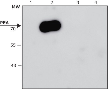
Western Blotting Analysis: A representative lot detected HSD17B10 in Western Blotting applications (Frackowiak, J., et. al. (2001). Brain Res. 907(1-2):44-53).
Immunohistochemistry Analysis: A 1:250-1:1,000 dilution from a representative lot detected HSD17B10 in human placenta and human breast cancer tissues.
Research Category
Neuroscience
품질
Evaluated by Western Blotting in HEK293T cell lysate.
Western Blotting Analysis: 0.5 µg/mL of this antibody detected HSD17B10 in 10 µg of HEK293T cell lysate.
표적 설명
~27 kDa observed; 26.92 kDa calculated. Uncharacterized bands may be observed in some lysate(s).
물리적 형태
Format: Purified
Protein G purified
Purified mouse monoclonal antibody IgG1 in buffer containing 0.1 M Tris-Glycine (pH 7.4), 150 mM NaCl with 0.05% sodium azide.
저장 및 안정성
Stable for 1 year at 2-8°C from date of receipt.
기타 정보
Concentration: Please refer to lot specific datasheet.
면책조항
Unless otherwise stated in our catalog or other company documentation accompanying the product(s), our products are intended for research use only and are not to be used for any other purpose, which includes but is not limited to, unauthorized commercial uses, in vitro diagnostic uses, ex vivo or in vivo therapeutic uses or any type of consumption or application to humans or animals.
