MABF977
Anti-ICOS/CD278 Antibody, clone C398.4A
clone C398.4A, from hamster(Armenian)
동의어(들):
Inducible T-cell costimulator, Activation-inducible lymphocyte immunomediatory molecule, CCLP, CD278, CD28 and CTLA-4-like protein, CD28-related protein 1, CRP-1, H4
About This Item
추천 제품
생물학적 소스
hamster (Armenian)
Quality Level
항체 형태
purified immunoglobulin
항체 생산 유형
primary antibodies
클론
C398.4A, monoclonal
종 반응성
mouse, human
종 반응성(상동성에 의해 예측)
rhesus macaque (based on 100% sequence homology)
기술
flow cytometry: suitable
immunocytochemistry: suitable
immunohistochemistry: suitable
immunoprecipitation (IP): suitable
동형
IgG
NCBI 수납 번호
UniProt 수납 번호
배송 상태
ambient
타겟 번역 후 변형
unmodified
유전자 정보
human ... ICOS(29851)
mouse ... Icos(54167)
관련 카테고리
일반 설명
특이성
면역원
애플리케이션
Flow Cytometry Analysis: A representative lot detected the surface expression of exogenously transfected human ICOS on the surface of murine L cell fibroblasts. Clone C398.4A and another ICOS mAb (clone F44) competed against each other for the the suface staining of PHA-activated human peripheral blood T-cells (Buonfiglio, D., et al. (2000). Eur. J. Immunol. 30(12):3463-3467).
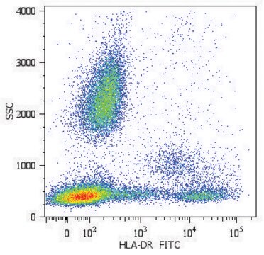
Flow Cytometry Analysis: A representative lot detected a greatly enhanced ICOS/CD278- (hpH4; human putative mouse H4 homolog) positive cell population among isolated human PBMCs following PHA stimulation (Buonfiglio, D., et al. (1999). Eur. J. Immunol. 29(9):2863-2874).
Flow Cytometry Analysis: A representative lot immunostained D10.G4.1 murine TH2 cells as well as activated, but not resting, T and B cells freshly isolated from mouse spleen (Redoglia, V., et al. (1996). Eur. J. Immunol. 26(11):2781-2789).
Immunocytochemistry Analysis: A representative lot immunostained H4 (ICOS/CD278) immunoreactivity co-capped with that of TCR mAb 3D3 on the surface of D10.G4.1 murine TH2 cells by immunofluorescence microscopy (Redoglia, V., et al. (1996). Eur. J. Immunol. 26(11):2781-2789).
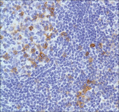
Immunohistochemistry Analysis: A representative lot immunostained the membrane of hpH4- (ICOS/CD278) positive lymphocytes in acetone-fixed frozen human reactive lymph node sections, while most neoplastic cells showed cytoplasmic hpH4 staining in frozen PTCL-U (peripheral T cell lymphoma, unspecified) and angioimmunoblastic T cell lymphoma sections (Buonfiglio, D., et al. (1999). Eur. J. Immunol. 29(9):2863-2874).
Immunoprecipitation Analysis: A representative lot immunoprecipitated and depleted ICOS from human T-cell lymphoma HuT 78 cell lysates (Buonfiglio, D., et al. (2000). Eur. J. Immunol. 30(12):3463-3467).
Immunoprecipitation Analysis: A representative lot immunoprecipitated H4 (ICOS/CD278) from PHA-activiated human peripheral blood (PB) T cells in a ~55-70 kDa glycosylated dimeric form (Buonfiglio, D., et al. (1999). Eur. J. Immunol. 29(9):2863-2874).
Immunoprecipitation Analysis: A representative lot immunoprecipitated H4 (ICOS/CD278) from D10.G4.1 murine TH2 cells in a glycosylated dimeric form (Redoglia, V., et al. (1996). Eur. J. Immunol. 26(11):2781-2789).
Inflammation & Immunology
품질
Flow Cytometry Analysis: 0.1 µg of this antibody detected ICOS/CD278 immunoreactivity on the surface of PHA-activated (5 µg/mL at 37°C for 72 hours) human PBMCs.
표적 설명
물리적 형태
저장 및 안정성
Handling Recommendations: Upon receipt and prior to removing the cap, centrifuge the vial and gently mix the solution. Aliquot into microcentrifuge tubes and store at -20°C. Avoid repeated freeze/thaw cycles, which may damage IgG and affect product performance.
기타 정보
면책조항
적합한 제품을 찾을 수 없으신가요?
당사의 제품 선택기 도구.을(를) 시도해 보세요.
Storage Class Code
12 - Non Combustible Liquids
WGK
WGK 2
Flash Point (°F)
Not applicable
Flash Point (°C)
Not applicable
시험 성적서(COA)
제품의 로트/배치 번호를 입력하여 시험 성적서(COA)을 검색하십시오. 로트 및 배치 번호는 제품 라벨에 있는 ‘로트’ 또는 ‘배치’라는 용어 뒤에서 찾을 수 있습니다.
자사의 과학자팀은 생명 과학, 재료 과학, 화학 합성, 크로마토그래피, 분석 및 기타 많은 영역을 포함한 모든 과학 분야에 경험이 있습니다..
고객지원팀으로 연락바랍니다.