추천 제품
생물학적 소스
mouse
Quality Level
항체 형태
purified immunoglobulin
항체 생산 유형
primary antibodies
클론
4-10-C9, monoclonal
종 반응성
mouse
기술
flow cytometry: suitable
immunocytochemistry: suitable
동형
IgG2aκ
NCBI 수납 번호
UniProt 수납 번호
배송 상태
wet ice
타겟 번역 후 변형
unmodified
유전자 정보
mouse ... Lag3(16768)
일반 설명
Lymphocyte activation gene 3 protein (UniProt Q61790; also known as CD223, LAG-3) is encoded by the Lag3 gene (Gene ID 16768) in murine species. LAG-3 is an inhibitory T-cell surface molecule that modulates T-cell activation and homeostasis. LAG-3 co-localizes with CD8 and CD4 upon TCR engagement and alters TCR signaling. LAG-3 suppresses homeostasis proliferation (HP) of both lymphocytes and some dendritic cell populations in vivo, and enhanced HP is observed in mice deficient in LAG-3. LAG-3 signaling is shown to negatively regulate STAT5 phosphorylation in activated T cells, and no enhancement of HP was seen upon LAG-3 blockade in mice deficient in both STAT5a and STAT5b. Murine LAG-3 is produced with a signal peptide sequence (a.a. 1-22), the removal of which yields the mature protein with a large extracellular region (a.a. 23-442), followed by a transmembrane segment (a.a. 443-463) and a cytoplasmic tail (a.a. 464-521). The extracellular portion contains a V-type Ig-like domain (a.a. 37-163), followed by three C2-type Ig-like domains (a.a. 165-246, 258-341, and 345-412) and elven tandem repeats of 2-amino acid E-X seqeunce at its C-terminal end (a.a. 493-518).
특이성
Clone 4-10-C9 immunostained surface LAG-3 on activated CD4+ T cells by targeting an extracellular epitope within the third and fourth Ig-like domains (D3/D4 domains; second and third C2-type Ig-like domains). Surface LAG-3 degradation by pronase treatment abolished cell surface staining (Woo, S.R., et al. (2010). Eur. J. Immunol. 40(6):1768-1777).
면역원
Epitope: Within D3/D4 domains.
Murine LAG-3-expressing mouse T-cell hybridoma.
애플리케이션
Flow Cytometry Analysis: 2.5 µg/mL from a representative lot detected surface LAG-3 immunoreactivity among the CD4+ and CD8+ populations of wild-type, but not Lag3-knockout, mouse splenocytes activated in vitro via CD3 cross-linking (Courtesy of Dario A. Vignali, Ph.D., University of Pittsburgh, PA, U.S.A.).
Flow Cytometry Analysis: A representative lot was fluorescently conjugated and detected an increased number of LAG-3-positive cells within the CD4+ and CD8+ populations of infiltrating lymphocytes (TILs) in tumors developed in mice exografted with murine B16 melanoma, MC38 colon adenocarcinoma, or Sa1N fibrosarcoma cells (Woo, S.R., et al. (2012). Cancer Res. 72(4): 917–927).
Flow Cytometry Analysis: A representative lot, pre-conjugated with Alexa Fluor™ 647, detected both surface and intracellular LAG-3 by immunofluorescent staining of non-permeabilized and permeabilized primary murine CD4+ T cells activated in vitro via CD3 & CD28 cross-linking by immobilized antibodies. Pronase treatment of cells prior to permeabilization abolished cell surface staining (Woo, S.R., et al. (2010). Eur. J. Immunol. 40(6):1768-1777).
Flow Cytometry Analysis: A representative lot, pre-conjugated with Alexa Fluor 647, detected a time-dependent recovery of cell surface LAG-3 immunoreactivity on activated murine CD4+ T cells after initial surface LAG-3 degradation by pronase treatment. Protein synthesis inhibitor cycloheximide (Cat. No. 239764) or protein transport inhibitor Brefeldin A (Cat. No. 203729) treatment partially blocked the recovery (Woo, S.R., et al. (2010). Eur. J. Immunol. 40(6):1768-1777).
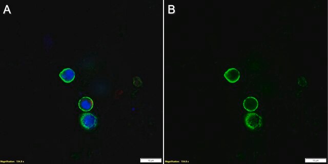
Immunocytochemistry Analysis: A representative lot detected both surface and intracellular LAG-3 by fluorescent immunocytochemistry staining of non-permeabilized and permeabilized primary murine CD4+ T cells activated in vitro via CD3 & CD28 cross-linking by immobilized antibodies. Pronase treatment of cells prior to permeabilization abolished cell surface staining (Woo, S.R., et al. (2010). Eur. J. Immunol. 40(6):1768-1777).
Immunocytochemistry Analysis: A representative lot detected intracellular LAG-3 immunoreactivity co-localized with those of the early and recycling endosome marker EEA1, as well as endosomal markers Rab11b and Rab27a by fluorescent immunocytochemistry staining of activated murine CD4+ T cells following pronase treatment and permeabilization (Woo, S.R., et al. (2010). Eur. J. Immunol. 40(6):1768-1777).
This Anti-LAG3 Antibody, clone 4-10-C9 is validated for use in Flow Cytometry, Immunocytochemistry for the detection of LAG3.
품질
Evaluated by Flow Cytometry in mouse splenocytes.
Flow Cytometry Analysis: 1 µg/mL of this antibody detected an induction of LAG-3-positive population in isolated mouse splenocytes following a 3-day 2 µg/mL Concanavalin A (Con A) stimulation.
Flow Cytometry Analysis: 1 µg/mL of this antibody detected an induction of LAG-3-positive population in isolated mouse splenocytes following a 3-day 2 µg/mL Concanavalin A (Con A) stimulation.
표적 설명
54.51/56.98 kDa (mature/pro-form) calculated.
물리적 형태
Format: Purified
법적 정보
ALEXA FLUOR is a trademark of Life Technologies
적합한 제품을 찾을 수 없으신가요?
당사의 제품 선택기 도구.을(를) 시도해 보세요.
Storage Class Code
12 - Non Combustible Liquids
WGK
WGK 1
시험 성적서(COA)
제품의 로트/배치 번호를 입력하여 시험 성적서(COA)을 검색하십시오. 로트 및 배치 번호는 제품 라벨에 있는 ‘로트’ 또는 ‘배치’라는 용어 뒤에서 찾을 수 있습니다.
Timo Eninger et al.
Proceedings of the National Academy of Sciences of the United States of America, 119(24), e2119804119-e2119804119 (2022-06-07)
Single-cell transcriptomics has revealed specific glial activation states associated with the pathogenesis of neurodegenerative diseases, such as Alzheimer’s and Parkinson’s disease. While these findings may eventually lead to new therapeutic opportunities, little is known about how these glial responses are
자사의 과학자팀은 생명 과학, 재료 과학, 화학 합성, 크로마토그래피, 분석 및 기타 많은 영역을 포함한 모든 과학 분야에 경험이 있습니다..
고객지원팀으로 연락바랍니다.