MABF2108
Anti-Chlamydial HSP60 Antibody, clone A57-B9
clone A57-B9, from mouse
동의어(들):
Chlamydia heat shock protein 60
로그인조직 및 계약 가격 보기
모든 사진(1)
About This Item
UNSPSC 코드:
12352203
eCl@ss:
32160702
NACRES:
NA.41
클론:
A57-B9, monoclonal
application:
ELISA
ICC
IF
WB
ICC
IF
WB
종 반응성:
bacteria, Chlamydia
기술:
ELISA: suitable
immunocytochemistry: suitable
immunofluorescence: suitable
western blot: suitable
immunocytochemistry: suitable
immunofluorescence: suitable
western blot: suitable
citations:
추천 제품
생물학적 소스
mouse
항체 형태
purified antibody
항체 생산 유형
primary antibodies
클론
A57-B9, monoclonal
종 반응성
bacteria, Chlamydia
포장
antibody small pack of 25 μg
기술
ELISA: suitable
immunocytochemistry: suitable
immunofluorescence: suitable
western blot: suitable
동형
IgG1κ
타겟 번역 후 변형
unmodified
일반 설명
Chlamydia trachomatis is a Gram-negative bacterium that is responsible to sexually transmitted diseases leading to pelvic inflammatory disease, ectopic pregnancy, infertility, and outbreaks of trachoma-associated blindness and lymphogranuloma venereum (LGV). Chlamydia trachomatis consists of eighteen different serological variants (serovars) that include a few subvariants. These are identified based on serological reactivity of the epitopes on their outer membrane. Intracellularly chlamydia replicates within a vacuole. Chlamydia infection is initiated with the expression of a chlamydial early gene product(s), which isolate the inclusion from the endocytic-lysosomal pathway and makes it fusogenic with sphingomyelin-containing exocytic vesicles. This change in vesicular interaction allows the delivery of the vacuole to the peri-Golgi region of the host cell. Antigens from all members of the Chlamydia genus display heat resistance and sensitivity to oxidation by sodium periodate. Clone A57-B9 detects Chlamydial HSP60 (GroEL). It reacts with the short peptide of 6 amino acids from the carboxyl terminal region of HSP60. HSP60 facilitates the correct folding of imported proteins and may prevent misfolding and promote the refolding and proper assembly of unfolded polypeptides generated under stress conditions.
특이성
Clone A57-B9 detects Chlamydial HSP60. It targets an epitope with in 144 amino acids in the carboxy terminal of HSP60.
면역원
Recombinant Chlamydia trachomatis serovar A HSP60 emulsified in complete Freund adjuvant.
애플리케이션
Anti-Chlamydial HSP60, clone A57-B9, Cat. No. MABF2108, is a mouse monoclonal antibody that detects Chlamydial HSP60 and has been tested for use in ELISA, Immunofluorescence, Immunocytochemistry, and Western Blotting.
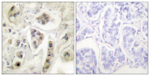
Immunocytochemistry Analysis: A representative lot detected Chlamydial HSP60 in Immunocytochemistry applications (Yuan, Y., et. al. (1992). Infect Immun. 60(6):2288-96).
Immunofluorescence Analysis: A representative lot detected Chlamydial HSP60 in Immunofluorescence applications (Southern, T., et. al. (2012). Clin Vaccine Immunol. 19(11):1864-9).
ELISA Analysis: A representative lot detected Chlamydial HSP60 in ELISA applications (Morrison, S.G., et. al. (2005). J Immunol. 175(11):7536-42).
Western Blotting Analysis: A representative lot detected chlamydial HSP60 in Western Blotting applications (Hechard, C., et. al. (2004). J Med Microbiol. 53(Pt 9):861-8; LaVerda, D., et. al. (1997). J Clin Microbiol. 35(5):1209-15; Southern, T., et. al. (2012). Clin Vaccine Immunol. 19(11):1864-9; Yuan, Y., et. al. (1992). Infect Immun. 60(6):2288-96).
Research Category
Inflammation & Immunology
Inflammation & Immunology
품질
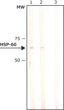
Evaluated by Western Blotting in lysates from HeLa cells infected with chlamydia trachomatis serovar L2 (LGV 434 L2) .
Western Blotting Analysis: 2 µg/mL of this antibody detected Chlamydial HSP60 in lysates from HeLa cells infected with Chlamydia trachomatis serovar L2 (LGV 434 L2) .
Western Blotting Analysis: 2 µg/mL of this antibody detected Chlamydial HSP60 in lysates from HeLa cells infected with Chlamydia trachomatis serovar L2 (LGV 434 L2) .
표적 설명
~60 kDa observed. Uncharacterized bands may be observed in some lysate(s).
물리적 형태
Format: Purified
Protein G purified
Purified mouse monoclonal antibody IgG1 in buffer containing 0.1 M Tris-Glycine (pH 7.4), 150 mM NaCl with 0.05% sodium azide.
저장 및 안정성
Stable for 1 year at 2-8°C from date of receipt.
기타 정보
Concentration: Please refer to lot specific datasheet.
면책조항
Unless otherwise stated in our catalog or other company documentation accompanying the product(s), our products are intended for research use only and are not to be used for any other purpose, which includes but is not limited to, unauthorized commercial uses, in vitro diagnostic uses, ex vivo or in vivo therapeutic uses or any type of consumption or application to humans or animals.
적합한 제품을 찾을 수 없으신가요?
당사의 제품 선택기 도구.을(를) 시도해 보세요.
Storage Class Code
12 - Non Combustible Liquids
WGK
WGK 1
Flash Point (°F)
Not applicable
Flash Point (°C)
Not applicable
시험 성적서(COA)
제품의 로트/배치 번호를 입력하여 시험 성적서(COA)을 검색하십시오. 로트 및 배치 번호는 제품 라벨에 있는 ‘로트’ 또는 ‘배치’라는 용어 뒤에서 찾을 수 있습니다.
자사의 과학자팀은 생명 과학, 재료 과학, 화학 합성, 크로마토그래피, 분석 및 기타 많은 영역을 포함한 모든 과학 분야에 경험이 있습니다..
고객지원팀으로 연락바랍니다.