추천 제품
생물학적 소스
mouse
Quality Level
항체 형태
purified immunoglobulin
항체 생산 유형
primary antibodies
클론
34, monoclonal
종 반응성
mouse, bovine, Xenopus, human, rat
종 반응성(상동성에 의해 예측)
ox (immunogen homology)
기술
flow cytometry: suitable
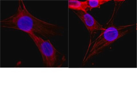
immunocytochemistry: suitable
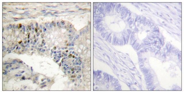
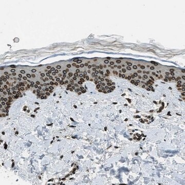
immunohistochemistry: suitable
western blot: suitable
동형
IgG1κ
NCBI 수납 번호
UniProt 수납 번호
배송 상태
wet ice
타겟 번역 후 변형
unmodified
유전자 정보
human ... H1F0(3005)
일반 설명
Histones are highly conserved proteins that serve as the structural scaffold for the organization of nuclear DNA into chromatin. The four core histones, H2A, H2B, H3, and H4, assemble into an octamer (2 molecules of each). Subsequently, 146 base pairs of DNA are wrapped around the octamer, forming a nucleosome. The linker histone, H1, interacts with linker DNA between nucleosomes and functions in the compaction of chromatin into 30nm chromatin fibers and higher order structures.
특이성
This antibody recognizes Histone H1°.
면역원
Recombinant protein corresponding to Ox liver Histone H1°
애플리케이션
Anti-Histone H1° Antibody, clone 34 is a highly specific mouse monoclonal antibody, that targets Histone H1 & has been tested in western blotting, ICC, IHC & Flow Cytometry.
Research Category
Epigenetics & Nuclear Function
Epigenetics & Nuclear Function
Research Sub Category
Histones
Histones
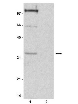
Western Blotting Analysis: A representative lot from an independent laboratory detected Histone H1° in Xenopus embryo tissue lysate (Seigneurin, D., et al. (1995). Int J Dev Biol. 39(4):597-603.; Fu, G., et al. (2003). Biol Reprod. 68(5):1569-1576.).
Immunocytochemistry Analyis: A representative lot from an independent laboratory detected Histone H1° in Xenopus unfertilized eggs and early embryos (Fu, G., et al. (2003). Biol Reprod. 68(5):1569-1576.; Adenot, P. G., et al. (2000). J Cell Sci. 113(Pt 16):2897-2907.).
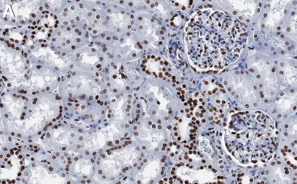
Immunohistochemistry Analysis: A representative lot from an independent laboratory detected Histone H1° in Xenopus embryo tissues (Grunwald, D., et al. (1995). Exp Cell Res. 218(2):586-595.).
Flow Cytometry Analyisis: A representative lot from an independent laboratory detected Histone H1° in FC (Grunwald, D., et al. (1999). Methods Mol Biol. 119:443-454.).
품질
Evaluated by Western Blotting in Jurkat cell lysate.
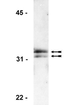
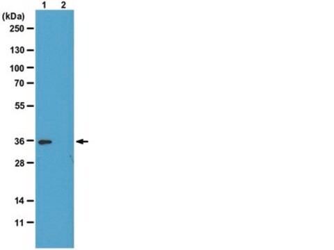
Western Blotting Analysis: 1 µg/mL of this antibody detected Histone H1° in 10 µg of Jurkat cell lysate.
Western Blotting Analysis: 1 µg/mL of this antibody detected Histone H1° in 10 µg of Jurkat cell lysate.
표적 설명
~30 kDa observed. Uncharacterized band(s) may be observed in some cell lysates.
물리적 형태
Format: Purified
Protein G Purified
Purified mouse monoclonal IgG1κ in buffer containing 0.1 M Tris-Glycine (pH 7.4), 150 mM NaCl with 0.05% sodium azide.
저장 및 안정성
Stable for 1 year at 2-8°C from date of receipt.
분석 메모
Control
Jurkat cell lysate
Jurkat cell lysate
기타 정보
Concentration: Please refer to the Certificate of Analysis for the lot-specific concentration.
면책조항
Unless otherwise stated in our catalog or other company documentation accompanying the product(s), our products are intended for research use only and are not to be used for any other purpose, which includes but is not limited to, unauthorized commercial uses, in vitro diagnostic uses, ex vivo or in vivo therapeutic uses or any type of consumption or application to humans or animals.
적합한 제품을 찾을 수 없으신가요?
당사의 제품 선택기 도구.을(를) 시도해 보세요.
Storage Class Code
12 - Non Combustible Liquids
WGK
WGK 1
Flash Point (°F)
Not applicable
Flash Point (°C)
Not applicable
시험 성적서(COA)
제품의 로트/배치 번호를 입력하여 시험 성적서(COA)을 검색하십시오. 로트 및 배치 번호는 제품 라벨에 있는 ‘로트’ 또는 ‘배치’라는 용어 뒤에서 찾을 수 있습니다.
Germaine Fu et al.
Biology of reproduction, 68(5), 1569-1576 (2003-02-28)
Oocytes and embryos of many species, including mammals, contain a unique linker (H1) histone, termed H1oo in mammals. It is uncertain, however, whether other H1 histones also contribute to the linker histone complement of these cells. Using immunofluorescence and radiolabeling
D Grunwald et al.
Experimental cell research, 218(2), 586-595 (1995-06-01)
It is known that a transition in the linker-histone variants takes place within chromatin during early development of Xenopus laevis; a cleavage-type H1 is replaced by the somatic type. Based on cytofluorimetric analysis of the distribution of the embryo cells
P G Adenot et al.
Journal of cell science, 113 ( Pt 16), 2897-2907 (2000-07-27)
A striking feature of early embryogenesis in a number of organisms is the use of embryonic linker histones or high mobility group proteins in place of somatic histone H1. The transition in chromatin composition towards somatic H1 appears to be
In situ analysis of chromatin proteins during development and cell differentiation using flow cytometry.
D Grunwald et al.
Methods in molecular biology (Clifton, N.J.), 119, 443-454 (2000-05-11)
Developmentally regulated chromatin acetylation and histone H1(0) accumulation.
Seigneurin, D, et al.
International Journal of Developmental Biology, 39, 597-603 (1995)
자사의 과학자팀은 생명 과학, 재료 과학, 화학 합성, 크로마토그래피, 분석 및 기타 많은 영역을 포함한 모든 과학 분야에 경험이 있습니다..
고객지원팀으로 연락바랍니다.
