추천 제품
생물학적 소스
mouse
Quality Level
항체 형태
purified antibody
항체 생산 유형
primary antibodies
클론
TwMab-1, monoclonal
분자량
calculated mol wt 127 kDa
observed mol wt ~127 kDa
정제법
using protein G
종 반응성
human
포장
antibody small pack of 100
기술
western blot: suitable
동형
IgG1κ
에피토프 서열
N-terminal half
단백질 ID 수납 번호
UniProt 수납 번호
저장 온도
2-8°C
특이성
Clone TwMab-1 is a mouse monoclonal antibody that detects Telomerase reverse transcriptase (TERT). It targets an epitope within 11 amino acids from the N-terminal half.
면역원
KLH-conjugated linear peptide corresponding to 11 amino acids from the N-terminal half of human Telomerase reverse transcriptase (TERT).
애플리케이션
Quality Control Testing
Isotype testing: Identity confirmation by Isotyping Test.
Isotyping Analysis: The identity of this monoclonal antibody is confirmed by isotyping test to be mouse IgG1k.
Tested Applications
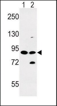
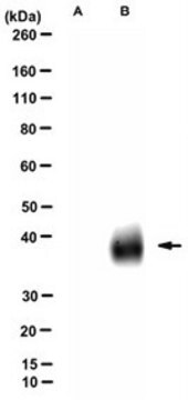
Western Blotting Analysis: A 1:100 dilution from a representative lot detected TERT in lysate from HEK293T cells with ectopic expression of hTERT (positive), but not in lysate from wild-type HEK293T cells (negative). ( Data courtesy of Dr. Kenkichi Masutomi, National Cancer Center Research Institute, Tokyo, Japan).
Note: Actual optimal working dilutions must be determined by end user as specimens, and experimental conditions may vary with the end user.
표적 설명
Telomerase reverse transcriptase (UniProt: O14746; also known as EC:2.7.7.49, HEST2, Telomerase catalytic subunit, Telomerase-associated protein 2, TP2) is encoded by the TERT (also known as EST2, TCS1, TRT) gene (Gene ID: 7015) in human. Telomerase is a ribonucleoprotein enzyme essential for the replication of chromosome termini in most eukaryotes. Telomerase reverse transcriptase (TERT) is the catalytic subunit of the telomerase responsible for adding TTAGGG repeats to the chromosome telomere ends. The catalytic cycle involves primer binding, primer extension and release of product once the template boundary has been reached or nascent product translocation followed by further extension. Cells with low or no telomerase expression lose telomere repeats during cell division, eventually resulting in cellular senescence. Most cancer cells, germ cells, and embryonic stem cells express high levels of telomerase, thus contributing to pluripotency and immortality. In addition to its telomere maintenance function, telomerase also has a pro-survival role in cellular resistance against DNA damage and ensuing apoptosis. Most cancer cells are highly proliferative and express high levels of nuclear telomerase activity. TERT is reported to shuttle between nuclear and cytoplasm and this shuttling is depended upon cell cycle, phosphorylation state, transformation, and DNA damage. TERT is also detected in the mitochondria where it helps prevent nuclear DNA damage by decreasing mitochondrial reactive oxygen species (ROS). Following exposure to H2O2 or -irradiation, cancer cells capable of excluding TERT from the nucleus display little or no DNA damage, while TERT nuclear retainment results in high DNA damage. Phosphorylation at tyrosine 707 under oxidative stress leads to translocation of TERT to the cytoplasm and reduces its anti-apoptotic activity and its dephosphorylation by SHP2/PTPN11 leads to its nuclear retention. Phosphorylation at serine 227 by AKT is reported to promote nuclear location. Phosphorylation of TERT at threonine 249 is reported to be a novel tumor biomarker of aggressive cancer with poor prognosis. (Ref.: Masuda, Y., et al. (2022). J. Pathol. 257(2); 172-185; Jacob, S., et al. (2008). J. Biol. Chem. 283(48); 33155-33161; Haendeler, J., et al. (2004). Circ. Res. 94(6); 768-775).
물리적 형태
Purified mouse monoclonal antibody IgG1 in buffer containing 0.1 M Tris-Glycine (pH 7.4), 150 mM NaCl with 0.05% sodium azide.
재구성
0.5 mg/mL. Please refer to guidance on suggested starting dilutions and/or titers per application and sample type.
저장 및 안정성
Stable for 1 year at +2°C to +8°C from date of receipt.
기타 정보
Concentration: Please refer to the Certificate of Analysis for the lot-specific concentration.
면책조항
Unless otherwise stated in our catalog or other company documentation accompanying the product(s), our products are intended for research use only and are not to be used for any other purpose, which includes but is not limited to, unauthorized commercial uses, in vitro diagnostic uses, ex vivo or in vivo therapeutic uses or any type of consumption or application to humans or animals.
적합한 제품을 찾을 수 없으신가요?
당사의 제품 선택기 도구.을(를) 시도해 보세요.
Storage Class Code
12 - Non Combustible Liquids
WGK
WGK 1
Flash Point (°F)
Not applicable
Flash Point (°C)
Not applicable
시험 성적서(COA)
제품의 로트/배치 번호를 입력하여 시험 성적서(COA)을 검색하십시오. 로트 및 배치 번호는 제품 라벨에 있는 ‘로트’ 또는 ‘배치’라는 용어 뒤에서 찾을 수 있습니다.
자사의 과학자팀은 생명 과학, 재료 과학, 화학 합성, 크로마토그래피, 분석 및 기타 많은 영역을 포함한 모든 과학 분야에 경험이 있습니다..
고객지원팀으로 연락바랍니다.
