추천 제품
생물학적 소스
mouse
Quality Level
항체 형태
ascites fluid
항체 생산 유형
primary antibodies
클론
6F5, monoclonal
종 반응성
mouse, human
기술
flow cytometry: suitable
immunocytochemistry: suitable
western blot: suitable
동형
IgG1
UniProt 수납 번호
배송 상태
wet ice
타겟 번역 후 변형
unmodified
유전자 정보
human ... SOD1(6647)
일반 설명
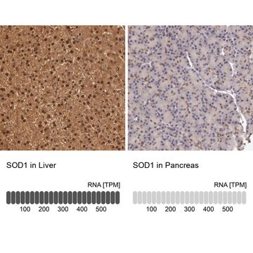
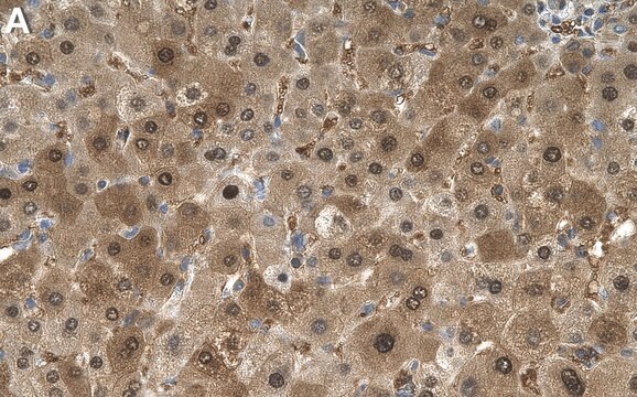
Superoxide dismutase 1 (SOD1) also known as Superoxide dismutase [Cu-Zn] to distinguish it from SOD2 which uses manganese as its cofactor, is the cytosolic form of the enzyme that catalyzes the breakdown of superoxide into oxygen and hydrogen peroxide and is our primary anti-oxidant combating enzyme. SOD1 performs its function with the help of co-factors and one protein in particular is vital to its proper functioning. Copper chaperone protein (CCS) is a metalloprotein that specifically delivers Cu to SOD1 and may activate SOD1 through direct insertion of the Cu cofactor. Interestingly, defects in SOD1 are the cause of a form of ALS (Amyotrophic Lateral Sclerosis or Lou Gehrig′s disease) and much research is centered on understanding how over-active or under-active SOD1 contributes to the disease. EMD-Millipore′s Anti-SOD1 mouse monoclonal antibody has been tested in western blot on HeLa, NIH3T3, A549 and A431 cell lysates and fluorescent immunocytochemistry on PANC1, SKBR-3 and 3T3-L1 cells in culture. The antibody has also been tested in flow cytometry on A431 cells in culture.
면역원
Purified recombinant fragment of human SOD1 expressed in E. Coli.
애플리케이션
Anti-SOD1 Antibody, clone 6F5 is a highly specific mouse monoclonal antibody, that targets Superoxide Dismutase & has been tested in western blotting, ICC & Flow Cytometry.
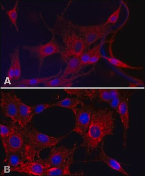
Immunocytochemistry Analysis: A 1:200-1,000 dilution from a representative lot detected SOD1 in PANC-1, SKBR-3, and 3T3-L1 cells.
Flow Cytometry Analysis: A 1:200-400 dilution from a representative lot detected SOD1 in non serum starved A431 cells.
Optimal working dilutions must be determined by end user.
Flow Cytometry Analysis: A 1:200-400 dilution from a representative lot detected SOD1 in non serum starved A431 cells.
Optimal working dilutions must be determined by end user.
Research Category
Apoptosis & Cancer
Apoptosis & Cancer
Research Sub Category
Apoptosis - Additional
Apoptosis - Additional
품질
Evaluated by Western Blotting in HeLa, NIH/3T3, A549, and A431 cell lysates.
Western Blotting Analysis: A 1:500-2,000 dilution of this antibody detected SOD1 in HeLa, NIH/3T3, A549, and A431 cell lysates.
Western Blotting Analysis: A 1:500-2,000 dilution of this antibody detected SOD1 in HeLa, NIH/3T3, A549, and A431 cell lysates.
표적 설명
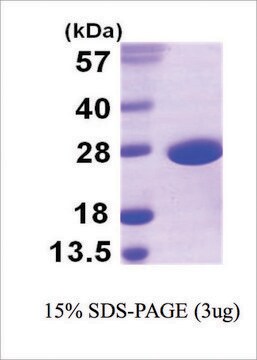
~18 kDa observed. Uncharacterized bands may appear in some lysate(s).
물리적 형태
Mouse monoclonal IgG1 ascitic fluid containing up to 0.1% sodium azide.
Unpurified
저장 및 안정성
Stable for 1 year at -20°C from date of receipt.
Handling Recommendations: Upon receipt and prior to removing the cap, centrifuge the vial and gently mix the solution. Aliquot into microcentrifuge tubes and store at -20°C. Avoid repeated freeze/thaw cycles, which may damage IgG and affect product performance.
Handling Recommendations: Upon receipt and prior to removing the cap, centrifuge the vial and gently mix the solution. Aliquot into microcentrifuge tubes and store at -20°C. Avoid repeated freeze/thaw cycles, which may damage IgG and affect product performance.
분석 메모
Control
HeLa, NIH/3T3, A549, and A431 cell lysates
HeLa, NIH/3T3, A549, and A431 cell lysates
면책조항
Unless otherwise stated in our catalog or other company documentation accompanying the product(s), our products are intended for research use only and are not to be used for any other purpose, which includes but is not limited to, unauthorized commercial uses, in vitro diagnostic uses, ex vivo or in vivo therapeutic uses or any type of consumption or application to humans or animals.
적합한 제품을 찾을 수 없으신가요?
당사의 제품 선택기 도구.을(를) 시도해 보세요.
Storage Class Code
12 - Non Combustible Liquids
WGK
nwg
Flash Point (°F)
Not applicable
Flash Point (°C)
Not applicable
시험 성적서(COA)
제품의 로트/배치 번호를 입력하여 시험 성적서(COA)을 검색하십시오. 로트 및 배치 번호는 제품 라벨에 있는 ‘로트’ 또는 ‘배치’라는 용어 뒤에서 찾을 수 있습니다.
Xiao-Dan Hao et al.
Scientific reports, 7(1), 13570-13570 (2017-10-21)
Keratoconus (KC) is a common degenerative corneal disease, and heredity plays a key role in its development. Although few genes are known to cause KC, a large proportion of disease-causing genes remain to be revealed. Here, we report the identification
자사의 과학자팀은 생명 과학, 재료 과학, 화학 합성, 크로마토그래피, 분석 및 기타 많은 영역을 포함한 모든 과학 분야에 경험이 있습니다..
고객지원팀으로 연락바랍니다.
