
추천 제품


생물학적 소스
mouse
Quality Level
항체 형태
purified antibody
항체 생산 유형
primary antibodies
클론
TA205, monoclonal
종 반응성
human
종 반응성(상동성에 의해 예측)
rabbit (based on 100% sequence homology)
기술
immunofluorescence: suitable
immunohistochemistry: suitable (paraffin)
immunoprecipitation (IP): suitable
inhibition assay: suitable
western blot: suitable
동형
IgG1κ
NCBI 수납 번호
UniProt 수납 번호
배송 상태
wet ice
타겟 번역 후 변형
unmodified
유전자 정보
human ... TLN1(7094)
일반 설명
Talin 1, also known as Talin 1, and encoded by the gene TLN1/KIAA1027, a large connecting protein located on the cytoplasmic side of most cellular projections. Talin appears to act as an anchoring protein connecting the cytoskeletal protein structures to the plasma membrane. Talin gets concentrated at regions of cell-substratum contact and in particular cells such as lymphocytes at regions of direct cell-cell contact. . Talin 1 binds with high affinity to vinculin and interacts with other proteins found in cellular projections including focal adhesion kinases (FAK) and intermediate filament proteins such as synemin and layilin. Additionally, Talin 1 because of its connections with cytoskeletal proteins talin is associated with many vital cell motion/growth processes from axon guidance to cell migration, muscle contraction, cell-cell interactions, immune responses and platelet activation to name but a few.
면역원
Epitope: N-terminus
GST-tagged recombinant protein corresponding to the N-terminus of human Talin 1.
애플리케이션
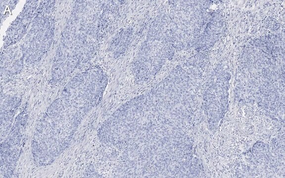
Immunohistochemistry Analysis: A 1:2,000 and a 1:500 dilution from a representative lot detected Talin 1 in human prostate cancer sections and human lung tissue, respectively.
Immunohistochemistry Analysis: A representative lot detected Talin 1 in MeOH fixed fibroblasts.
Inhibition Analysis: A representative lot from an independent laboratory disrupts actin stress fibers and focal adhesions, and inhibits cell motility when microinjected into human fibroblasts (Bolton, S. J., et al. (1997). Cell Motil Cytoskeleton. 36(4): 363-376.).
Immunoprecipitation Analysis: A representative lot from an independent laboratory immunoprecipitated Talin 1 from shCTRL and shLRP-1-c1 carcinoma cell lysates (Dedieu, S., et al. (2008). Mol Cell Biol. 28(9):2980-2995.).
Immunoprecipitation Analysis: A representative lot from an independent laboratory immunoprecipitated Talin 1 from shCTRL and shLRP-1 cell lysates (Langlois, B., et al. (2010). PLoS One. 5(7):e11584.).
Immunoprecipitation/Western Blotting Analysis: A representative lot from an independent laboratory immunoprecipitated Talin 1 from PC3-2 cell lysates. Immunoprecipitated Talin 1 was then detected in Western Blotting (Trerotola, M. et al. (2012). J Cell Physiol. 227(11): 3670-3676.).
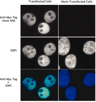
Immunofluorescence Analysis: A representative lot from an independent laboratory detected Talin 1 in macrophages (Love, D. C., et al. (1998). Exp Parasitol. 88(3):161-171.).
Immunofluorescence Analysis: A representative lot from an independent laboratory determined that the level of disruption in talin-containing focal complexes is inhibited in LRP-1-silenced carcinoma cells (Dedieu, S., et al. (2008). Mol Cell Biol. 28(9):2980-2995.).
Immunofluorescence Analysis: A representative lot from an independent laboratory detected Talin-1 in shCTRL and shLRP-1 transfected cells (Langlois, B., et al. (2010). PLoS One. 5(7):e11584.).
Immunohistochemistry Analysis: A representative lot detected Talin 1 in MeOH fixed fibroblasts.
Inhibition Analysis: A representative lot from an independent laboratory disrupts actin stress fibers and focal adhesions, and inhibits cell motility when microinjected into human fibroblasts (Bolton, S. J., et al. (1997). Cell Motil Cytoskeleton. 36(4): 363-376.).
Immunoprecipitation Analysis: A representative lot from an independent laboratory immunoprecipitated Talin 1 from shCTRL and shLRP-1-c1 carcinoma cell lysates (Dedieu, S., et al. (2008). Mol Cell Biol. 28(9):2980-2995.).
Immunoprecipitation Analysis: A representative lot from an independent laboratory immunoprecipitated Talin 1 from shCTRL and shLRP-1 cell lysates (Langlois, B., et al. (2010). PLoS One. 5(7):e11584.).
Immunoprecipitation/Western Blotting Analysis: A representative lot from an independent laboratory immunoprecipitated Talin 1 from PC3-2 cell lysates. Immunoprecipitated Talin 1 was then detected in Western Blotting (Trerotola, M. et al. (2012). J Cell Physiol. 227(11): 3670-3676.).
Immunofluorescence Analysis: A representative lot from an independent laboratory detected Talin 1 in macrophages (Love, D. C., et al. (1998). Exp Parasitol. 88(3):161-171.).
Immunofluorescence Analysis: A representative lot from an independent laboratory determined that the level of disruption in talin-containing focal complexes is inhibited in LRP-1-silenced carcinoma cells (Dedieu, S., et al. (2008). Mol Cell Biol. 28(9):2980-2995.).
Immunofluorescence Analysis: A representative lot from an independent laboratory detected Talin-1 in shCTRL and shLRP-1 transfected cells (Langlois, B., et al. (2010). PLoS One. 5(7):e11584.).
Research Category
Cell Structure
Cell Structure
Research Sub Category
Cytoskeleton
Cytoskeleton
This Anti-Talin 1 Antibody, clone TA205, N-Term, Ascites Free is validated for use in western blotting, IHC (Paraffin), IP, inhibition & immunofluorescence for the detection of Talin 1.
품질
Evaluated by Western Blotting in HeLa cell lysate.
Western Blotting Analysis: 1 µg/mL of this antibody detected Talin 1 in 10 µg of HeLa cell lysate.
Western Blotting Analysis: 1 µg/mL of this antibody detected Talin 1 in 10 µg of HeLa cell lysate.
표적 설명
~270 kDa observed
물리적 형태
Format: Purified
Protein G Purified
Purified mouse monoclonal IgG1κ in buffer containing PBS without preservatives.
저장 및 안정성
Stable for 1 year at -20°C from date of receipt.
Handling Recommendations: Upon receipt and prior to removing the cap, centrifuge the vial and gently mix the solution. Aliquot into microcentrifuge tubes and store at -20°C. Avoid repeated freeze/thaw cycles, which may damage IgG and affect product performance.
Handling Recommendations: Upon receipt and prior to removing the cap, centrifuge the vial and gently mix the solution. Aliquot into microcentrifuge tubes and store at -20°C. Avoid repeated freeze/thaw cycles, which may damage IgG and affect product performance.
기타 정보
Concentration: Please refer to the Certificate of Analysis for the lot-specific concentration.
면책조항
Unless otherwise stated in our catalog or other company documentation accompanying the product(s), our products are intended for research use only and are not to be used for any other purpose, which includes but is not limited to, unauthorized commercial uses, in vitro diagnostic uses, ex vivo or in vivo therapeutic uses or any type of consumption or application to humans or animals.
적합한 제품을 찾을 수 없으신가요?
당사의 제품 선택기 도구.을(를) 시도해 보세요.
Storage Class Code
12 - Non Combustible Liquids
WGK
WGK 2
Flash Point (°F)
Not applicable
Flash Point (°C)
Not applicable
시험 성적서(COA)
제품의 로트/배치 번호를 입력하여 시험 성적서(COA)을 검색하십시오. 로트 및 배치 번호는 제품 라벨에 있는 ‘로트’ 또는 ‘배치’라는 용어 뒤에서 찾을 수 있습니다.
자사의 과학자팀은 생명 과학, 재료 과학, 화학 합성, 크로마토그래피, 분석 및 기타 많은 영역을 포함한 모든 과학 분야에 경험이 있습니다..
고객지원팀으로 연락바랍니다.