CBL251
Anti-Epithelial Specific Antigen Antibody, clone VU-1D9
clone VU-1D9, from mouse
동의어(들):
Epithelial cell adhesion molecule, Ep-CAM, Adenocarcinoma-associated antigen, Cell surface glycoprotein Trop-1, Epithelial cell surface antigen, Epithelial glycoprotein, EGP, Epithelial glycoprotein 314, EGP314, hEGP314, KS 1/4 antigen, KSA, Major gastro
로그인조직 및 계약 가격 보기
모든 사진(3)
About This Item
UNSPSC 코드:
12352203
eCl@ss:
32160702
NACRES:
NA.41
추천 제품
일반 설명
Epithelial specific antigen, also known as epithelial cell adhesion molecule (Ep-CAM) and epithelial glycoprotein (EGP), may act as an interaction molecule between intestinal epithelial cells (IECs) and intraepithelial lymphocytes (IELs) at the mucosal epithelium for providing immunological barrier as a first line of defense against mucosal infection. Epithelial specific antigen plays a role in embryonic stem cells proliferation and differentiation, and up-regulates the expression of FABP5, MYC, and cyclins A and E. Epithelial specific antigen is highly and selectively expressed by undifferentiated embryonic stem cells (ESCs). Protein levels rapidly diminish as soon as ESCs differentiate. Also expressed in most epithelial cell membranes and on the surface of adenocarcinoma.
면역원
NCI-H69 whole cells
애플리케이션
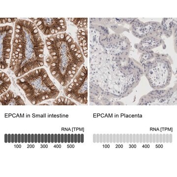
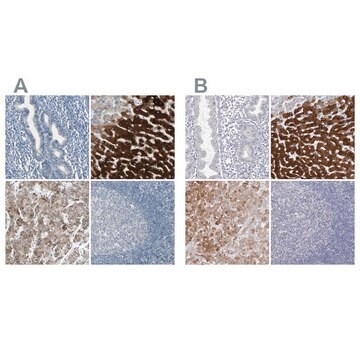
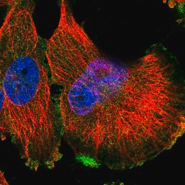
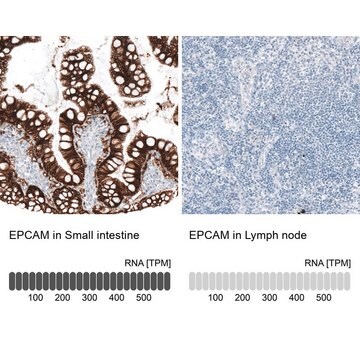
Anti-Epithelial Specific Antigen Antibody, clone VU-1D9 is a highly specific mouse monoclonal antibody, that targets Epithelial Cell Marker & has been tested in western blotting, ICC & IHC.
Immunocytochemistry Analysis: A 1:500 dilution from a representative lot detected Epithelial Specific Antigen in A431 cells.
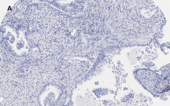
Immunohistochemistry Analysis: A 1:50 dilution from a representative lot detected Epithelial Specific Antigen in human prostate adenocarcinoma cells.
Immunohistochemistry Analysis: A 1:50 dilution from a representative lot detected Epithelial Specific Antigen in human prostate adenocarcinoma cells.
Research Category
Cell Structure
Cell Structure
Research Sub Category
ECM Proteins
ECM Proteins
품질
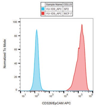
Evaluated by Western Blotting in MCF7 cell lyate.
Western Blotting Analysis: 1 µg/mL of this antibody detected Epithelial Specific Antigen in 10 µg of MCF7 cell lyate.
Western Blotting Analysis: 1 µg/mL of this antibody detected Epithelial Specific Antigen in 10 µg of MCF7 cell lyate.
표적 설명
~40 kDa observed. The calculated molecular weight of this protein is 35 kDa; however, this protein will run higher due to glycosylation.
물리적 형태
Format: Purified
Protein G Purified
Purified mouse monoclonal IgG1κ in buffer containing PBS with 0.05% sodium azide.
저장 및 안정성
Stable for 1 year at 2-8°C from date of receipt.
분석 메모
Control
MCF7 cell lyasate
MCF7 cell lyasate
기타 정보
Concentration: Please refer to the Certificate of Analysis for the lot-specific concentration.
면책조항
Unless otherwise stated in our catalog or other company documentation accompanying the product(s), our products are intended for research use only and are not to be used for any other purpose, which includes but is not limited to, unauthorized commercial uses, in vitro diagnostic uses, ex vivo or in vivo therapeutic uses or any type of consumption or application to humans or animals.
적합한 제품을 찾을 수 없으신가요?
당사의 제품 선택기 도구.을(를) 시도해 보세요.
Storage Class Code
10 - Combustible liquids
WGK
WGK 2
Flash Point (°F)
Not applicable
Flash Point (°C)
Not applicable
시험 성적서(COA)
제품의 로트/배치 번호를 입력하여 시험 성적서(COA)을 검색하십시오. 로트 및 배치 번호는 제품 라벨에 있는 ‘로트’ 또는 ‘배치’라는 용어 뒤에서 찾을 수 있습니다.
자사의 과학자팀은 생명 과학, 재료 과학, 화학 합성, 크로마토그래피, 분석 및 기타 많은 영역을 포함한 모든 과학 분야에 경험이 있습니다..
고객지원팀으로 연락바랍니다.