추천 제품
생물학적 소스
rabbit
Quality Level
항체 형태
unpurified
항체 생산 유형
primary antibodies
클론
polyclonal
종 반응성
mouse, human
종 반응성(상동성에 의해 예측)
rat (based on high sequence homology)
기술
immunofluorescence: suitable
immunohistochemistry: suitable (paraffin)
western blot: suitable
동형
IgG
NCBI 수납 번호
UniProt 수납 번호
배송 상태
ambient
타겟 번역 후 변형
unmodified
유전자 정보
human ... PTGES(9536)
일반 설명
Prostaglandin E synthase (UniProt: O14684; also known as EC:5.3.99.3, Microsomal glutathione S-transferase 1-like 1, MGST1-L1, Microsomal prostaglandin E synthase 1, MPGES-1, p53-induced gene 12 protein) is encoded by the PTGES gene (Gene ID: 9536) in human. mPGES -1 is a multi-pass membrane protein that is integral membrane, inducible and glutathione-dependent terminal synthase in the prostanoid biosynthesis pathway and acts downstream cyclooxygenases (COX) catalyzing the oxidoreduction of prostaglandin endoperoxide H2 (PGH2) to prostaglandin E2 (PGE2). Functionally, mPGES-1 is coupled to COX-2 and the induction of these two enzymes by pro-inflammatory cytokines, growth factors or endotoxin leads to a surge of PGE2 synthesis by inflammatory cells. It is strongly expressed with COX-2 in synovial fibroblasts and macrophages. In cases of osteoarthritis, mPGES-1 and COX-2 are strongly expressed in the chondrocytes and the synovial lining cells. mPGES-1 also play a role in the pathogenesis of ischemic stroke and many neurodegenerative diseases. In Alzheimer disease brains higher expression of both COX-2 and mPGES-1 is observed. Higher expression of mPGES has also been reported in several cancers and this higher expression has been correlated with vascular invasion and worse prognosis in colorectal cancer. A549 and DU145 xenografts lacking mPGES-1 display significantly reduced growth rates. (Ref.: Korotkova, M., and Jakobsson, PJ (2014). Basic Clin. Pharmacol. Toxicol. 114, 64-69).
특이성
This polyclonal antibody specifically detects mPGES-1 in human.
면역원
His-tagged full-length recombinant human microsomal prostaglandin E synthase.
애플리케이션
Detect Prostaglandin E synthase using this rabbit polyclonal Anti-mPTGES-1, Cat. No. ABS2192 that has been tested in Immunohistochemistry (Paraffin), Immunohistochemistry (Frozen), Immunofluorescence, and Western Blotting.
Immunohistochemistry Analysis: A 1:5,000 dilution from a representative lot detected mPTGES-1 in human tonsil and human placenta tissue.
Western Blotting Analysis: A representative lot detected mPTGES-1 in human chondrocytes (Tuure, L., et. al. (2015). Scand J Rheumatol. 44(1):74-9) and wild-type mixed glial cultures (Straccia, M., et. al. (2013). Glia. 61(10):1607-19).
Immunofluorescence Analysis: A representative lot detected mPTGES-1 in Wild-type mixed glial cultures (Tuure, L., et. al. (2015). Scand J Rheumatol. 44(1):74-9).
Western Blotting Analysis: A representative lot detected mPTGES-1 in PNE inhibitedmPGES-1proteinexpressioninIL-1 -stimulated SK-N-SH cells (Olajide, O.A., et. al. (2014). J Ethnopharmacol. 152(2):377-83).
Immunohistochemistry Analysis: A representative lot detected mPTGES-1 in fresh frozen brain tissue (Tuure, L., et. al. (2015). Scand J Rheumatol. 44(1):74-9).
품질
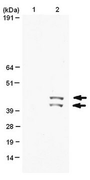
Evaluated by Western Blotting in recombinant microsomal Prostaglandin E synthase 1 protein.
Western Blotting Analysis: A 1:10,000 dilution of this antibody detected mPTGES-1 in recombinant microsomal Prostaglandin E synthase 1 protein.
Western Blotting Analysis: A 1:10,000 dilution of this antibody detected mPTGES-1 in recombinant microsomal Prostaglandin E synthase 1 protein.
표적 설명
~17 kDa observed; 17.10 kDa calculated. Uncharacterized bands may be observed in some lysate(s).
물리적 형태
Format: Unpurified
Rabbit polyclonal antiserum without azide.
기타 정보
Concentration: Please refer to lot specific datasheet.
적합한 제품을 찾을 수 없으신가요?
당사의 제품 선택기 도구.을(를) 시도해 보세요.
Storage Class Code
10 - Combustible liquids
WGK
WGK 1
Flash Point (°F)
Not applicable
Flash Point (°C)
Not applicable
시험 성적서(COA)
제품의 로트/배치 번호를 입력하여 시험 성적서(COA)을 검색하십시오. 로트 및 배치 번호는 제품 라벨에 있는 ‘로트’ 또는 ‘배치’라는 용어 뒤에서 찾을 수 있습니다.
자사의 과학자팀은 생명 과학, 재료 과학, 화학 합성, 크로마토그래피, 분석 및 기타 많은 영역을 포함한 모든 과학 분야에 경험이 있습니다..
고객지원팀으로 연락바랍니다.