추천 제품
생물학적 소스
rabbit
Quality Level
항체 형태
affinity isolated antibody
항체 생산 유형
primary antibodies
클론
polyclonal
정제법
affinity chromatography
종 반응성
human
종 반응성(상동성에 의해 예측)
bovine (based on 100% sequence homology)
기술
immunofluorescence: suitable
immunohistochemistry: suitable (paraffin)
western blot: suitable
NCBI 수납 번호
UniProt 수납 번호
배송 상태
wet ice
타겟 번역 후 변형
unmodified
유전자 정보
human ... APOE(348)
일반 설명
Apolipoprotein E (UniProt P02649; also known as Apo-E) is encoded by the APOE (also known as AD2, LDLCQ5, LPG) gene (Gene ID 348) in human. Apolipoprotein E (ApoE) is found in the chylomicron and Intermediate-density lipoprotein (IDLs) and is essential for the normal catabolism of triglyceride-rich lipoprotein constituents. ApoE is initially produced with a signal peptide sequence (a.a. 1-18), the removal of which yields the 299 a.a. mature protein. Three additional natural variants (E2, E3, and E4) exist due to polymorphisms. E4 has an allele frequency of approximately 14 percent and has been implicated in atherosclerosis and various CNS disorders, including Alzheimer′s disease (AD). ApoE4 is highly susceptible to proteolysis, which impairs its role in cholesterol transport and beta-amyloid removal in the CNS. Various ApoE4 fragments (14–20 kDa) have been identified in the AD brain. The C-terminal portion of apoE has been implicated in binding to beta-amyloid and is localized to plaques, while the N-terminal domain preferentially localizes within neurofibrillary tangles (NFTs) in the AD brain.
특이성
Specifically recognizes the ApoE4 N-terminal fragment(s) generated by a proteolytic cleavage between Asp172 and Leu173 of ApoE4. Does not recognize full-length ApoE3 and ApoE4, nor ApoE4 fragments with C-termi other than D172.
면역원
Epitope: C-terminus
Linear peptide corresponding to the C-terminal sequence of human ApoE4 fragment nApoECF (Asp172).
애플리케이션
Anti-ApoE4 Fragment nApoECF Antibody (Asp172) is an antibody against ApoE4 Fragment nApoECF for use in Immunohistochemistry (Paraffin), Western Blotting, Immunofluorescence.
Immunohistochemistry Analysis: A 1:1,000 dilution from a representative lot detected little ApoE4 fragment nApoECF (Asp172) immunoreactivity in normal human brain tissue.
Western Blotting Analysis: 1.0 µg/mL from a representative lot detected ApoE4 fragment nApoECF (Asp172) in 10 µg of human Alzheimer′s diseased brain tissue lysate.
Immunofluorescence Analysis: A representative lot detected a strong nApoECF (n-terminal ApoE4 Cleavage Fragment) immunoreactivity co-localized with that of PHF-1 within Pick bodies of area CA1 by dual-fluorescent immunohistochemistry using free-floating hippocampus tissue sections from a Pick′s disease patient (Rohn, T.T., et al. (2013). PLoS One. 8(12):e80180).
Immunofluorescence Analysis: A representative lot detected nApoECF (n-terminal ApoE4 Cleavage Fragment) immunoreactivity co-localized with that of cleaved Tau (Asp421; Cat. No. 36-017) within Pick bodies of area CA1 by dual-fluorescent immunohistochemistry using free-floating hippocampus tissue sections from a Pick′s disease patient (Rohn, T.T., et al. (2013). PLoS One. 8(12):e80180).
Immunofluorescence Analysis:A representative lot detected the the nApoECF (n-terminal ApoE4 Cleavage Fragment) immunoreactivity co-localized with the PHF-1-positive neurofibrillary tangles (NFTs) in the frontal cortex of Alzheimer′s diseased brains by dual-fluorescent immunohistochemistry using formic acid-treated free-floating sections (Rohn, T.T., et al. (2012). Brain Res. 1475:106-115).
Immunohistochemistry Analysis: A representative lot detected a strong nApoECF (n-terminal ApoE4 Cleavage Fragment) immunoreactivity within Pick bodies of area CA1 among 4 out of 5 Pick′s disease patients-derived free-floating hippocampus specimens (Rohn, T.T., et al. (2013). PLoS One. 8(12):e80180).
Immunohistochemistry Analysis: A representative lot detected the specific association of the nApoECF (n-terminal ApoE4 Cleavage Fragment) immunoreactivity with the neurofibrillary tangles (NFTs), but not within the Abeta-positive senile plaques, in the frontal cortex of Alzheimer′s diseased brains using formic acid-treated free-floating sections (Rohn, T.T., et al. (2012). Brain Res. 1475:106-115).
Western Blotting Analysis: A representative lot detected the 18 kDa nApoECF (n-terminal ApoE4 Cleavage Fragment; a.a. 1-172) present in the preparations of bacterially expressed human ApoE4, but not the full-length ApoE4 itself or the caspse-3-cleaved 16 kDa ApoE4 fragment (Rohn, T.T., et al. (2012). Brain Res. 1475:106-115).
Note: This antibody will detect any ApoE fragments with Asp172 at the C-terminus. This antibody was raised against a hydrophobic immunogen sequence and therefore exhibits high affinity toward hydrophobic membrane surface. Multiple banding pattern and overall high background are expected when using this antibody for Western blotting applications, especially when employing tissue samples.
Research Category
Neuroscience
Neuroscience
Research Sub Category
Neurodegenerative Diseases
Neurodegenerative Diseases
품질
Evaluated by Immunohistochemistry in Alzheimer′s diseased human brain tissue.
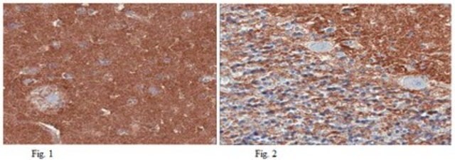
Immunohistochemistry Analysis: A 1:250 dilution of this antibody detected ApoE4 fragment nApoECF (Asp172) in Alzheimer′s diseased human brain tissue.
Immunohistochemistry Analysis: A 1:250 dilution of this antibody detected ApoE4 fragment nApoECF (Asp172) in Alzheimer′s diseased human brain tissue.
표적 설명
~18 kDa calculated
물리적 형태
Affinity purified
Purified rabbit polyclonal antibody in buffer containing 0.1 M Tris-Glycine (pH 7.4), 150 mM NaCl with 0.05% sodium azide.
저장 및 안정성
Stable for 1 year at 2-8°C from date of receipt.
기타 정보
Concentration: Please refer to lot specific datasheet.
면책조항
Unless otherwise stated in our catalog or other company documentation accompanying the product(s), our products are intended for research use only and are not to be used for any other purpose, which includes but is not limited to, unauthorized commercial uses, in vitro diagnostic uses, ex vivo or in vivo therapeutic uses or any type of consumption or application to humans or animals.
적합한 제품을 찾을 수 없으신가요?
당사의 제품 선택기 도구.을(를) 시도해 보세요.
Storage Class Code
12 - Non Combustible Liquids
WGK
WGK 1
Flash Point (°F)
Not applicable
Flash Point (°C)
Not applicable
시험 성적서(COA)
제품의 로트/배치 번호를 입력하여 시험 성적서(COA)을 검색하십시오. 로트 및 배치 번호는 제품 라벨에 있는 ‘로트’ 또는 ‘배치’라는 용어 뒤에서 찾을 수 있습니다.
Troy T Rohn et al.
Brain research, 1475, 106-115 (2012-08-21)
Although the risk factor for harboring the apolipoprotein E4 (apoE4) allele in late-onset Alzheimer's disease (AD) is well known, the mechanism by which apoE4 contributes to AD pathogenesis has yet to be clarified. Preferential cleavage of the ApoE4 isoform relative
Troy T Rohn et al.
PloS one, 8(12), e80180-e80180 (2013-12-07)
Although the risk factor for apolipoprotein E (apoE) polymorphism in Alzheimer's disease (AD) has been well described, the role that apoE plays in other neurodegenerative diseases, including Pick's disease, is not well established. To examine a possible role of apoE
자사의 과학자팀은 생명 과학, 재료 과학, 화학 합성, 크로마토그래피, 분석 및 기타 많은 영역을 포함한 모든 과학 분야에 경험이 있습니다..
고객지원팀으로 연락바랍니다.


