
추천 제품


생물학적 소스
rabbit
Quality Level
항체 형태
purified antibody
항체 생산 유형
primary antibodies
클론
polyclonal
종 반응성
mouse
제조업체/상표
Chemicon®
기술
immunohistochemistry: suitable (paraffin)
NCBI 수납 번호
UniProt 수납 번호
배송 상태
wet ice
타겟 번역 후 변형
unmodified
유전자 정보
human ... OPN1LW(5956)
일반 설명
Long-wave-sensitive opsin 1/Medium-wave-sensitive opsin 1 (UniProt: P04000/P04001; also known as Red cone photoreceptor pigment/Green cone photoreceptor pigment, Red-sensitive opsin/ Green-sensitive opsin, ROP/GOP) are encoded by the OPN1LW/OPN1MW (also known as RCP/GCP) genes (Gene ID: 5956/2652) in human. The full range of color discrimination in humans is based on the presence and function of three cone photoreceptors. Each cone type possesses a photo-sensitive pigment-protein complex consisting of 11-cis retinal and a unique opsin protein that gives sensitivity in the short (S cone, peak sensitivity about 420 nm), middle (M cone, peak sensitivity about 530 nm with polymorphism), and long (L cone, peak sensitivity about 560 nm with polymorphism) wavelengths of the light spectrum. Opsins are multi-pass membrane proteins that belongs to the G-protein coupled receptor 1 family. They consist of four extracellular, 7 helical, and four cytoplasmic domains. Genes for the three types of cone opsins and the rod photoreceptor rhodopsin gene seem to be homologous with varying amounts of conservation. Strongest conservation is between the middle (green) and long (red) wavelength sensitive pigments on the X chromosome, suggesting a relatively recent duplication/divergence event. The S cone (blue) opsin seems to have a stronger conservation with rhodopsin. Cone photoreceptor distribution in humans is dominated by the M and L cone pigments. Mutations in OPN1MW and OPN1LW genes are known to cause color blindness that is characterized by a dichromasy in which red and green are confused, without loss of luminance or shift or shortening of the spectrum. Some mutations also lead to cone dystrophy leading to progressive degeneration of the cone photoreceptor with some preservation of rod function. (Ref.: Neitz, M., and Neitz, J. (2000). Arch. Ophthalmol. 118(5); 691-700).
특이성
Mouse. Reactivity with other species has not been determined
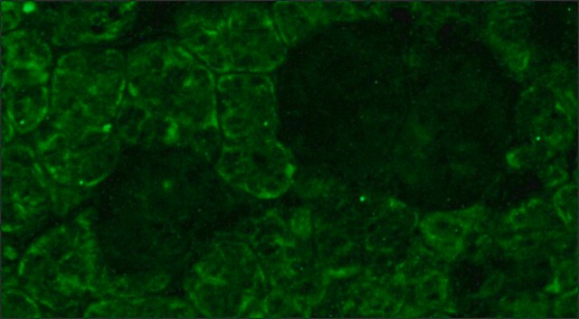
This rabbit polyclonal antibody detects Red-sensitive opsin/ Green-sensitive opsins.
면역원
Full-length, recombinant human red/green opsin.
Recombinant human red/green opsin.
애플리케이션
Anti-Opsin Red/Green, Cat. No. AB5405, is a rabbit polyclonal antibody that detects Opsin Red/Green and is tested for use in Immunohistochemistry (Paraffin).
품질
Evaluated by Immunohistochemistry (Paraffin) in Mouse retina tissue sections.Immunohistochemistry (Paraffin) Analysis: A 1:250 dilution of this antibody detected Opsin Red/Green in Mouse retina tissue sections.
물리적 형태
Format: Purified
Purified rabbit polyclonal antibody in buffer containing 0.02 M phosphate buffer, pH 7.6, 0.25 M NaCl, and 0.1% sodium azide.
저장 및 안정성
Recommended storage: +2°C to +8°C.
기타 정보
Concentration: Please refer to the Certificate of Analysis for the lot-specific concentration.
법적 정보
CHEMICON is a registered trademark of Merck KGaA, Darmstadt, Germany
적합한 제품을 찾을 수 없으신가요?
당사의 제품 선택기 도구.을(를) 시도해 보세요.
Storage Class Code
12 - Non Combustible Liquids
WGK
WGK 2
Flash Point (°F)
Not applicable
Flash Point (°C)
Not applicable
시험 성적서(COA)
제품의 로트/배치 번호를 입력하여 시험 성적서(COA)을 검색하십시오. 로트 및 배치 번호는 제품 라벨에 있는 ‘로트’ 또는 ‘배치’라는 용어 뒤에서 찾을 수 있습니다.
Analysis of gene function in the retina.
Takahiko Matsuda,Constance L Cepko
Methods in Molecular Biology null
Rescue of retinal degeneration by intravitreally injected adult bone marrow-derived lineage-negative hematopoietic stem cells.
Otani, Atsushi, et al.
The Journal of Clinical Investigation, 114, 765-774 (2004)
In vivo and in vitro development of S- and M-cones in rat retina.
Arango-Gonzalez, B; Szabo, A; Pinzon-Duarte, G; Lukats, A; Guenther, E; Kohler, K
Investigative Ophthalmology & Visual Science null
The electroretinogram (ERG) of a diurnal cone-rich laboratory rodent, the Nile grass rat (Arvicanthis niloticus).
Gilmour, Gregory S, et al.
Vision Research, 48, 2723-2731 (2008)
Severe retinal degeneration caused by a novel rhodopsin mutation.
Liu H, Wang M, Xia CH, Du X, Flannery JG, Ridge KD, Beutler B, Gong X
Investigative Ophthalmology & Visual Science null
자사의 과학자팀은 생명 과학, 재료 과학, 화학 합성, 크로마토그래피, 분석 및 기타 많은 영역을 포함한 모든 과학 분야에 경험이 있습니다..
고객지원팀으로 연락바랍니다.